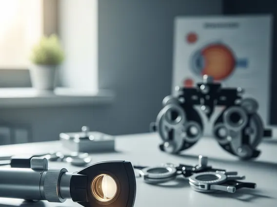
Direct Ophthalmoscopy
Direct ophthalmoscopy is a fundamental clinical procedure used to examine the interior structures of the eye, particularly the retina, optic disc, macula, and retinal blood vessels. It provides a direct, magnified view of the posterior segment, aiding in the diagnosis and monitoring of various ocular and systemic conditions.
Key Takeaways
- Direct ophthalmoscopy is a non-invasive examination of the eye’s internal structures using a handheld ophthalmoscope.
- It helps detect and monitor various eye conditions, including glaucoma, diabetic retinopathy, and macular degeneration.
- The procedure involves systematically viewing the optic disc, retinal blood vessels, and macula.
- It is a crucial diagnostic tool for early detection and management of both ocular and systemic diseases.
What is Direct Ophthalmoscopy?
Direct ophthalmoscopy is a common and essential diagnostic technique in ophthalmology and general medicine. It involves using a handheld instrument called a direct ophthalmoscope to illuminate and magnify the internal structures of the eye, primarily the fundus (retina, optic disc, macula, and retinal blood vessels). This method provides an upright, real image of the fundus, allowing clinicians to assess the health of these vital components directly. The examination is non-invasive and provides a highly magnified view, typically 15 times, making it invaluable for detailed observation of the posterior segment.
What is Direct Ophthalmoscopy Used For?
Direct ophthalmoscopy is a versatile diagnostic tool with numerous applications in healthcare, serving as a cornerstone for detecting and monitoring a wide range of ocular and systemic diseases. The Direct ophthalmoscopy indications and benefits include its ability to identify early signs of conditions that might otherwise go unnoticed until more advanced stages. By visualizing the retina and optic nerve, practitioners can assess for abnormalities indicative of various health issues.
It is primarily used to:
- Detect retinal diseases such as diabetic retinopathy, which can lead to vision loss if untreated.
- Identify signs of glaucoma by observing changes in the optic disc, such as cupping.
- Diagnose macular degeneration, a leading cause of vision loss in older adults, by examining the macula.
- Assess for hypertensive retinopathy, where high blood pressure affects retinal blood vessels.
- Screen for optic nerve abnormalities, including optic neuritis or papilledema, which can indicate neurological conditions.
- Monitor the progression of existing eye conditions and the effectiveness of treatments.
This examination is crucial for both routine eye check-ups and the investigation of specific symptoms, contributing significantly to preventive care and timely intervention.
How to Do a Direct Ophthalmoscopy Exam
Performing a direct ophthalmoscopy exam requires skill and practice to obtain a clear view of the fundus. The Direct ophthalmoscopy technique guide outlines a systematic approach to ensure a thorough and effective examination. The procedure is typically performed in a darkened room to facilitate pupil dilation and enhance visibility. The patient is usually seated, and the examiner positions themselves close to the patient, aligning their eye with the ophthalmoscope and the patient’s eye.
Here are the general steps involved in performing a direct ophthalmoscopy exam:
- Patient Preparation: Explain the procedure to the patient. Dim the room lights. If necessary, administer dilating eye drops, though many examinations can be performed through an undilated pupil.
- Ophthalmoscope Setup: Turn on the ophthalmoscope light. Set the diopter wheel to zero or to compensate for your own refractive error.
- Initial Approach: Stand about 15 inches (38 cm) away from the patient, at an angle of 15-20 degrees to their line of sight. Shine the light into the pupil to observe the red reflex.
- Fundus Examination: Slowly move closer to the patient, maintaining the red reflex. As you get closer, you will begin to see the retinal structures. Adjust the diopter wheel to bring the structures into sharp focus.
- Systematic Scan: Examine the optic disc first, noting its color, margins, and the presence of any cupping. Then, follow the retinal blood vessels peripherally. Finally, ask the patient to look directly into the light to examine the macula and fovea, which are crucial for central vision.
- Documentation: Record all findings, including any abnormalities observed in the optic disc, blood vessels, macula, and retina.
This systematic approach helps ensure that all critical areas of the fundus are thoroughly inspected, allowing for accurate diagnosis and monitoring.