3d Crt
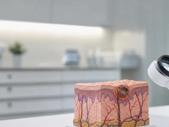
3D Conformal Radiation Therapy (3D CRT) represents a significant advancement in cancer treatment, utilizing sophisticated imaging and planning to precisely deliver radiation doses. This technique aims to maximize tumor eradication while minimizing damage to surrounding healthy tissues.

Key Takeaways
- 3D Conformal Radiation Therapy (3D CRT) is a radiation technique that shapes radiation beams to conform to the precise shape of a tumor.
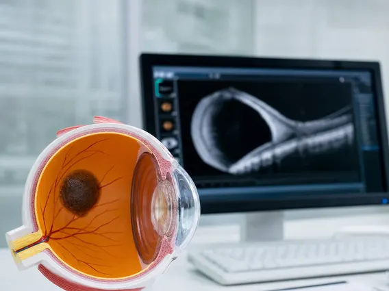
- It uses advanced imaging (CT, MRI, PET) to create a detailed 3D map of the tumor and surrounding organs.
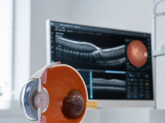
- Specialized display systems, often referred to as 3D CRT monitors, are crucial for visualizing and planning the complex radiation dose distribution.
- The technology designs multiple radiation beams that converge on the tumor, sparing healthy tissue.
- 3D CRT was a foundational step towards more advanced radiation therapies and remains a valuable treatment option for various cancers.
What is 3D CRT Technology?
3D Conformal Radiation Therapy (3D CRT) is a type of external beam radiation therapy that uses computers and advanced imaging to create a detailed, three-dimensional map of a tumor. This technology allows radiation oncologists to shape radiation beams to precisely match the contours of the tumor, delivering a high dose of radiation to cancerous cells while sparing nearby healthy tissues and organs. The primary goal of 3D CRT technology is to improve treatment effectiveness and reduce side effects by optimizing the radiation dose distribution.
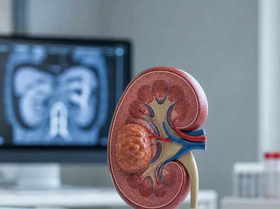
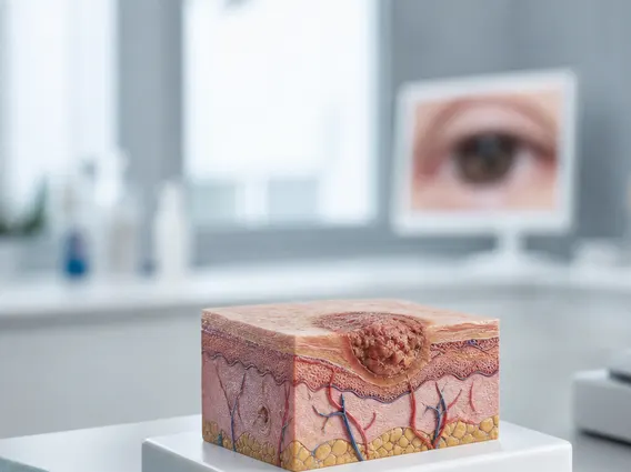
This approach involves a meticulous planning process. Patients undergo imaging scans, typically computed tomography (CT) scans, but often supplemented with magnetic resonance imaging (MRI) or positron emission tomography (PET) scans, to define the exact size, shape, and location of the tumor. Using this data, a treatment planning system generates a virtual 3D model, enabling the medical team to design radiation fields that “conform” to the tumor’s shape. This precision is crucial for treating various cancers, including those of the prostate, lung, and brain.
How do 3D CRT Monitors Work?
In the context of 3D Conformal Radiation Therapy, the term “3D CRT monitors” refers to the specialized display systems and software interfaces used by radiation oncologists, dosimetrists, and radiation therapists. These high-resolution displays are integral to the precise planning and delivery of radiation treatment. They enable clinicians to visualize complex anatomical structures, tumor volumes, and proposed radiation dose distributions in three dimensions. The functionality of these monitors is critical for ensuring the accuracy and safety of the therapy.
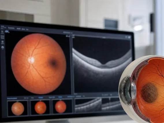
The explanation of 3D CRT displays involves understanding their role in translating complex imaging data into actionable treatment plans. These systems work by processing vast amounts of imaging data from CT, MRI, and PET scans. The software reconstructs this data into detailed 3D models of the patient’s body, highlighting the tumor and surrounding organs at risk. Clinicians use these specialized displays to:
- Contour Target Volumes: Precisely outline the tumor (Gross Tumor Volume, Clinical Target Volume, Planning Target Volume).
- Identify Organs at Risk (OARs): Delineate healthy organs that need to be protected from radiation.
- Design Beam Arrangements: Virtually position multiple radiation beams to converge on the tumor while avoiding OARs.
- Evaluate Dose Distribution: Analyze how the radiation dose is distributed throughout the patient’s body, ensuring adequate coverage of the tumor and minimal exposure to healthy tissues.
This visualization and interactive planning capability on specialized displays is fundamental to the success of 3D CRT.
History and Evolution of 3D CRT Technology
The development of 3D Conformal Radiation Therapy marked a significant paradigm shift in oncology, moving beyond two-dimensional treatment planning. Early radiation therapy relied on 2D X-ray images, making it challenging to precisely target tumors and protect surrounding healthy tissues. The advent of computed tomography (CT) in the 1970s and 1980s provided the foundational imaging capability needed for 3D planning. This allowed clinicians to visualize patient anatomy in cross-sections, which could then be reconstructed into a three-dimensional model.
The true breakthrough for the History and evolution of 3D CRT came with the integration of powerful computing capabilities and sophisticated treatment planning software in the late 1980s and early 1990s. This allowed for the design of radiation beams that could be shaped to conform to the tumor’s exact geometry. Before 3D CRT, radiation fields were typically simple squares or rectangles, leading to more collateral damage to healthy tissue. The ability to use custom-shaped blocks or multi-leaf collimators (MLCs) to sculpt the radiation beams was a revolutionary step. According to the American Society for Radiation Oncology (ASTRO), 3D CRT became widely adopted in the 1990s, significantly improving the therapeutic ratio—the balance between tumor control and normal tissue toxicity. This technology laid the groundwork for even more advanced techniques such as Intensity-Modulated Radiation Therapy (IMRT) and Stereotactic Body Radiation Therapy (SBRT), which further refine dose delivery and conformality.