3 Tesla Magnetic Resonance Imaging
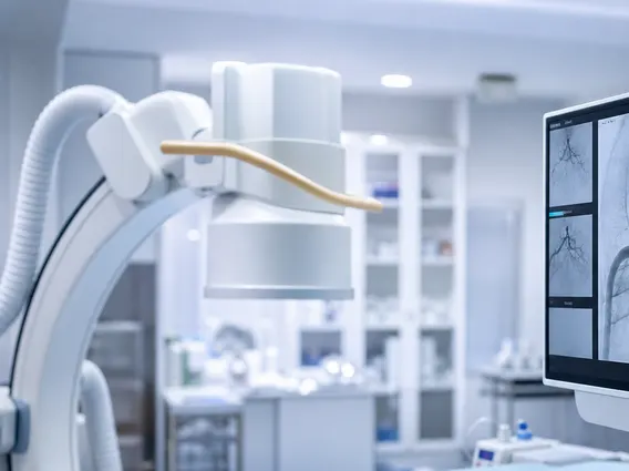
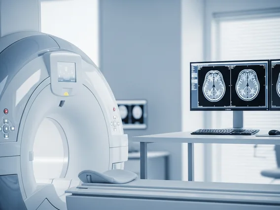
3 Tesla Magnetic Resonance Imaging (3T MRI) represents an advanced diagnostic tool that utilizes a powerful magnetic field to produce highly detailed images of organs, soft tissues, bone, and virtually all other internal body structures. This technology plays a crucial role in modern medicine by aiding in the accurate diagnosis and monitoring of various conditions.
Key Takeaways
- 3 Tesla Magnetic Resonance Imaging (3T MRI) uses a magnetic field twice as strong as standard 1.5T MRI, leading to superior image clarity.
- The stronger magnetic field in 3T MRI enhances signal detection, allowing for faster scans and more detailed anatomical and functional insights.
- 3T MRI benefits include higher spatial resolution, improved signal-to-noise ratio, and better visualization of subtle abnormalities, particularly in neurological and musculoskeletal imaging.
- When considering 3 tesla mri vs 1.5 tesla mri, 3T offers enhanced detail but may present louder noise and specific considerations for patients with certain metallic implants.
- Potential 3T MRI risks are generally similar to those of lower-field MRI, primarily related to the strong magnetic field’s interaction with metallic objects or implants.
What is 3 Tesla Magnetic Resonance Imaging (3T MRI)?
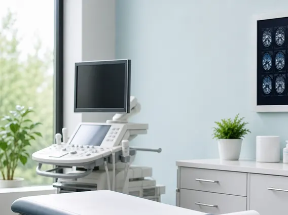
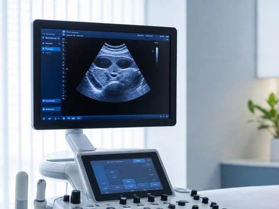
3 Tesla Magnetic Resonance Imaging (3T MRI) refers to a sophisticated medical imaging technique that employs a magnetic field strength of 3 Tesla, which is twice as powerful as the standard 1.5 Tesla MRI systems commonly found in clinical settings. This increased magnetic field strength is central to its ability to generate exceptionally clear and detailed images of the body’s internal structures. By interacting with the hydrogen atoms within the body, the powerful magnet and radiofrequency pulses allow for the creation of high-resolution cross-sectional images without using ionizing radiation.
The “Tesla” unit measures magnetic field strength, with higher values indicating a more potent magnetic field. The enhanced field of a 3T MRI scanner allows for a stronger signal from the body’s tissues, translating into superior image quality and diagnostic precision. This capability is particularly advantageous for visualizing intricate anatomical details and subtle pathological changes that might be less apparent on lower-field MRI scans.
How 3 Tesla MRI Works and Its Clinical Advantages
The fundamental principle behind how 3 tesla mri works involves harnessing the magnetic properties of hydrogen atoms, which are abundant in the body’s water molecules. When a patient is placed inside the powerful 3 Tesla magnet, these hydrogen protons align with the magnetic field. Radiofrequency pulses are then briefly emitted, knocking the protons out of alignment. When these pulses are turned off, the protons relax back into alignment, releasing energy signals that are detected by the MRI scanner. The stronger 3 Tesla magnetic field generates a more robust signal, which significantly improves the clarity and detail of the resulting images.
The clinical advantages of 3T MRI are substantial, offering enhanced diagnostic capabilities across various medical specialties. The primary 3T MRI benefits stem from its ability to produce images with higher spatial resolution and an improved signal-to-noise ratio (SNR). This allows radiologists to visualize smaller structures and subtle abnormalities with greater confidence. Key benefits include:
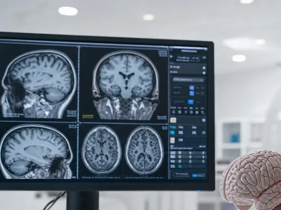
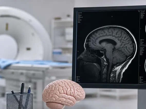
- Superior Image Resolution: Enables clearer visualization of fine anatomical details, crucial for diagnosing conditions in complex areas like the brain, spine, and joints.
- Faster Scan Times: The stronger signal often allows for quicker acquisition of images, reducing patient discomfort and motion artifacts.
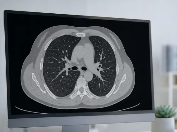
- Enhanced Contrast: Provides better differentiation between healthy and diseased tissues, which is particularly valuable in oncology for tumor detection and characterization.
- Advanced Functional Imaging: Facilitates specialized techniques such as functional MRI (fMRI) for brain activity mapping and diffusion tensor imaging (DTI) for white matter tractography.
These advantages make 3T MRI an invaluable tool for diagnosing neurological disorders, musculoskeletal injuries, and various cancers, offering more precise information for treatment planning.
Comparing 3T MRI to 1.5T MRI and Associated Risks
The comparison between 3 tesla mri vs 1.5 tesla mri highlights distinct differences in performance and application. While both are highly effective diagnostic tools, the 3T system offers a significant leap in image quality due to its stronger magnetic field. This translates into finer detail and better tissue contrast, which can be critical for certain diagnoses. However, the increased field strength also comes with specific considerations regarding patient safety and comfort.
| Feature | 1.5 Tesla MRI | 3 Tesla MRI |
|---|---|---|
| Magnetic Field Strength | Standard (1.5 Tesla) | High (3 Tesla) |
| Image Resolution | Good | Excellent (Higher spatial resolution) |
| Signal-to-Noise Ratio (SNR) | Good | Superior (Higher SNR) |
| Scan Time | Standard | Potentially faster for certain sequences |
| Noise Level | Moderate | Louder (Requires hearing protection) |
| Artifacts from Implants | Lower potential | Higher potential (More sensitive to metallic objects) |
| Specific Applications | General diagnostic imaging | Detailed neurological, musculoskeletal, and vascular imaging |
While 3T MRI offers significant diagnostic advantages, it is important to be aware of potential 3T MRI risks. The stronger magnetic field can interact more intensely with metallic objects and implants within or on the body. Patients with pacemakers, certain cochlear implants, aneurysm clips, or other metallic foreign bodies may not be eligible for an MRI, or specific precautions must be taken. The increased magnetic field can also lead to more pronounced heating of some metallic implants, although safety protocols are strictly followed to mitigate this. Additionally, the stronger magnetic gradients can generate louder acoustic noise during the scan, necessitating the use of earplugs or headphones for patient comfort and hearing protection. As with any medical procedure, a thorough screening process is conducted to ensure patient safety before undergoing a 3T MRI.