Hdr
Hdr refers to High Dynamic Range, a technology that significantly enhances the visual quality of images by expanding the contrast ratio and color accuracy. In medical imaging, this capability is crucial for improving diagnostic precision and clinical assessment.

Key Takeaways
- HDR (High Dynamic Range) is a technology that broadens the spectrum of light and color captured and displayed in images.
- It works by combining multiple exposures or using specialized sensors to record a wider range of luminance values.
- In medical imaging, HDR enhances the visualization of subtle tissue differences and anatomical structures.
- The improved contrast and detail provided by HDR can lead to more accurate diagnoses and better treatment planning.
What is HDR (High Dynamic Range)?
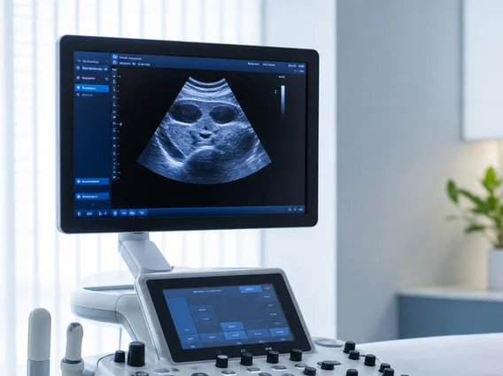
High Dynamic Range (HDR) refers to an imaging technology designed to reproduce a greater range of luminosity and color than standard dynamic range (SDR) imaging. This expanded range allows for the simultaneous display of very bright and very dark areas within a single image, preserving detail in both extremes. In the context of medical imaging, HDR explained means capturing and presenting a more faithful representation of the subtle variations in tissue density and structure, which are often critical for accurate diagnosis. Traditional imaging methods can struggle to adequately display both highly dense and less dense areas in the same view without losing information in one extreme. HDR overcomes this limitation by significantly increasing the contrast ratio and color depth, enabling clinicians to perceive nuances that might otherwise be overlooked.
How Does HDR Work?
The fundamental principle behind HDR involves capturing and processing a much wider spectrum of light intensity than conventional imaging. This is typically achieved through several methods:
- Multiple Exposure Fusion: One common technique involves taking several images of the same scene at different exposure levels (e.g., underexposed, normally exposed, and overexposed). These images are then algorithmically combined to create a single HDR image that retains detail in both the brightest highlights and the deepest shadows.
- Specialized Sensors: Some advanced imaging systems incorporate sensors capable of capturing a wider dynamic range in a single shot. These sensors often have a higher bit depth, allowing them to record more distinct levels of brightness and color information.
- Tone Mapping: Once the high dynamic range data is acquired, it undergoes a process called tone mapping. This process intelligently compresses the extensive luminance range into a displayable format while preserving perceived contrast and detail. This ensures that the image, despite having a vast internal dynamic range, can be effectively viewed on a standard display or printed, optimizing visual clarity for medical professionals.
Benefits of HDR in Medical Imaging
The integration of HDR technology into medical imaging offers substantial advantages for patient care and diagnostic accuracy. The primary benefits of HDR stem from its ability to provide a more comprehensive and detailed visual representation of internal structures. This enhanced clarity is particularly valuable in fields such as radiology, pathology, and endoscopy.
Key advantages include:
- Improved Visualization of Subtle Details: HDR allows radiologists and clinicians to discern minute differences in tissue density, vascular structures, or lesion characteristics that might be obscured by limited contrast in SDR images. This can be crucial for early detection and precise localization of abnormalities.
- Enhanced Diagnostic Confidence: By presenting a more complete picture with greater detail in both bright and dark regions, HDR images reduce the ambiguity often associated with standard imaging. This leads to higher confidence in diagnostic interpretations and treatment planning.
- Better Assessment of Disease Progression: The consistent and accurate representation of tissue characteristics over time, facilitated by HDR, aids in monitoring disease progression or response to treatment with greater precision.
- Reduced Need for Repeat Scans: With clearer initial images, the necessity for follow-up or repeat scans due to insufficient image quality can be reduced, improving patient experience and operational efficiency.
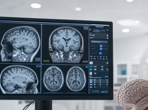
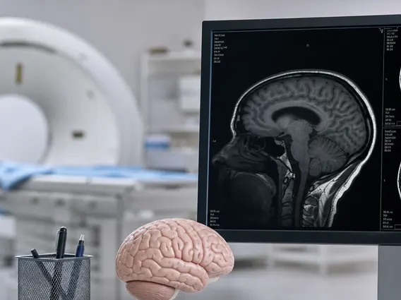
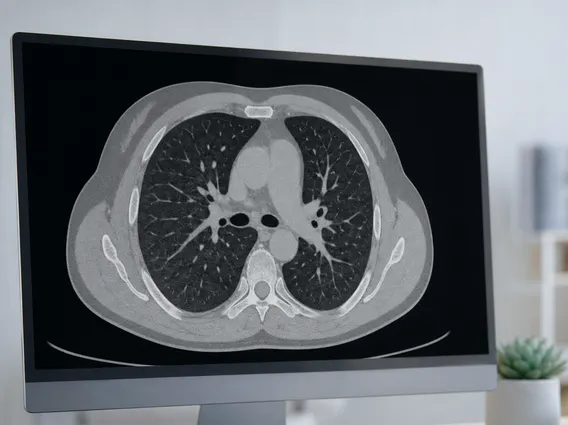
For example, in computed tomography (CT) scans, HDR can help differentiate between various soft tissues and bone structures more effectively, especially in complex anatomical areas. Similarly, in magnetic resonance imaging (MRI), it can enhance the visibility of subtle pathological changes within organs. While specific global statistics on HDR adoption in medical imaging are still emerging, the general trend in medical technology emphasizes advancements that improve diagnostic accuracy and patient outcomes, aligning perfectly with HDR’s capabilities.