Nmri
Nmri represents a significant advancement in medical diagnostics, offering unique insights into the human body’s internal structures and functions. This article explores the fundamental aspects of Nmri, from its core definition to the intricate mechanisms that underpin its operation.
Key Takeaways
- Nmri is a sophisticated medical imaging technique used for detailed visualization of internal body structures.
- Its primary purpose is to aid in the diagnosis, monitoring, and treatment planning of various medical conditions.
- Nmri operates by utilizing specific physical principles to generate high-resolution images without the use of ionizing radiation.
- The technology provides clinicians with non-invasive tools to assess tissue health and identify abnormalities.
What is Nmri and Its Purpose?
Nmri refers to a non-invasive medical imaging technique that provides highly detailed images of organs, soft tissues, bone, and virtually all other internal body structures. Unlike X-rays or CT scans, Nmri does not use ionizing radiation, making it a safer option for repeated examinations, especially for vulnerable populations or long-term monitoring. The Nmri meaning and explanation centers on its ability to differentiate between various tissue types based on their unique physical properties, offering clinicians a powerful diagnostic tool that can reveal subtle abnormalities often missed by other imaging modalities.
The primary purpose of Nmri is to assist in the diagnosis, monitoring, and management of a wide range of medical conditions across almost every part of the body. Its versatility makes it indispensable in several medical specialties. For instance, it is particularly valuable for:
- Neurological conditions: Detecting brain tumors, strokes, multiple sclerosis, and spinal cord injuries by clearly visualizing brain tissue and nerve pathways.
- Musculoskeletal issues: Accurately visualizing joint damage, ligament tears, cartilage degeneration, and bone infections, which are often difficult to assess with X-rays alone.
- Cardiovascular diseases: Assessing heart structure and function, identifying blockages in blood vessels, and evaluating damage from heart attacks, providing critical information for cardiac care.
- Oncology: Staging cancers, monitoring tumor growth, and evaluating treatment response in various organs, offering a non-invasive way to track disease progression.
How Nmri Technology Works
The sophisticated process of Nmri technology relies on fundamental principles of physics to generate its detailed images. At its core, Understanding Nmri principles involves the interaction of strong magnetic fields and radio waves with the body’s hydrogen atoms, which are abundant in water and fat. This interaction creates signals that are then translated into comprehensive visual data.
Here’s a simplified breakdown of how Nmri works:
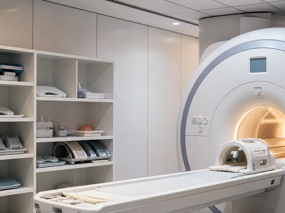
- Patient Positioning and Magnetic Field: The patient is carefully positioned inside a large, powerful cylindrical magnet. This magnet creates a strong, uniform magnetic field that causes the protons within the body’s hydrogen atoms (primarily from water molecules) to align themselves with the direction of this field.
- Radiofrequency Pulse Application: A brief, specific radiofrequency current is then pulsed through the patient. This energy temporarily excites the aligned protons, knocking them out of their alignment with the main magnetic field. The duration and frequency of these pulses are precisely controlled.
- Signal Emission and Relaxation: When the radiofrequency pulse is turned off, the excited protons relax and gradually return to their original alignment with the main magnetic field. As they relax, they release the absorbed energy in the form of radio signals. The rate at which different tissues relax varies significantly, and this difference is key to image formation.
- Signal Detection and Image Reconstruction: Receiver coils within the Nmri scanner detect these emitted radio signals. A powerful computer then processes these complex signals, using advanced algorithms to reconstruct them into detailed, cross-sectional images. These images can be rendered in multiple planes (e.g., axial, sagittal, coronal), providing clinicians with a comprehensive, three-dimensional understanding of the internal anatomy and pathology.
This intricate interplay of magnetic fields, radio waves, and sophisticated computing allows Nmri to produce high-resolution images that are crucial for accurate diagnosis and effective treatment planning, offering unparalleled soft tissue contrast.