Digital Breast Tomosynthesis
Digital Breast Tomosynthesis (DBT) represents a significant advancement in breast imaging technology, offering a more detailed and accurate method for breast cancer detection. This innovative technique provides a clearer view of breast tissue, enhancing diagnostic capabilities compared to traditional methods.
Key Takeaways
- Digital Breast Tomosynthesis (DBT) is an advanced 3D imaging technique for breast cancer detection.
- It works by taking multiple low-dose X-ray images from different angles, which are then reconstructed into a 3D volume.
- DBT offers improved cancer detection rates and reduced false positives compared to 2D mammography.
- The technology helps radiologists distinguish between overlapping breast tissues and actual abnormalities more effectively.
- DBT is increasingly becoming a preferred method for breast cancer screening and diagnosis.
What is Digital Breast Tomosynthesis (DBT)?
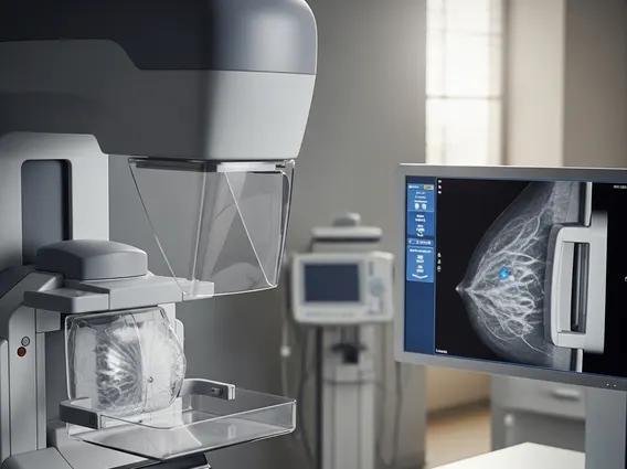
Digital Breast Tomosynthesis (DBT) refers to an advanced form of breast imaging that uses a low-dose X-ray system and computer reconstructions to create three-dimensional (3D) images of the breast. Unlike traditional 2D mammography, which captures a single flat image, DBT acquires multiple images as the X-ray tube moves in an arc over the breast. These individual images are then synthesized by a computer to produce a layered, highly detailed view of the breast tissue. This capability allows radiologists to scroll through the breast tissue slice by slice, similar to reviewing pages in a book, making it easier to detect abnormalities and differentiate them from overlapping normal tissue. The primary goal of DBT is to improve the early detection of breast cancer, particularly in women with dense breast tissue, where tumors can be obscured in 2D images.
How Digital Breast Tomosynthesis Works
The process of how breast tomosynthesis works involves several key steps to generate its detailed 3D images. During a DBT examination, the patient’s breast is positioned and compressed, similar to a traditional mammogram, to ensure optimal image quality and reduce radiation dose. The X-ray tube then moves in a sweeping arc over the compressed breast, capturing a series of low-dose images from various angles. Typically, around 11 to 25 projection images are acquired over a few seconds. A powerful computer then processes these individual images. Using complex algorithms, the computer reconstructs the data into a series of thin, high-resolution cross-sectional “slices” of the breast tissue. Each slice can be as thin as one millimeter. This layered reconstruction allows radiologists to examine the breast tissue in fine detail, effectively separating overlapping structures that might otherwise obscure a lesion or mimic an abnormality in a 2D image. This method significantly enhances the visibility of subtle lesions and microcalcifications, which are often early indicators of breast cancer.
Digital Breast Tomosynthesis vs. Traditional Mammography
Comparing digital breast tomosynthesis vs mammogram highlights the significant advancements DBT offers over its traditional counterpart. While both methods utilize X-rays to image breast tissue, their approaches to image acquisition and presentation differ fundamentally. Traditional mammography captures a single 2D image of the breast from a fixed angle, which can lead to issues with tissue overlap, making it challenging to distinguish between normal dense tissue and actual cancerous lesions. This can result in both missed cancers and false positives, potentially leading to unnecessary callbacks for additional imaging.
Digital Breast Tomosynthesis, on the other hand, provides a 3D view, effectively minimizing the problem of overlapping tissue. This leads to several benefits of digital breast tomosynthesis, including improved cancer detection rates and a reduction in false positives. The ability to view the breast in thin slices allows radiologists to more confidently identify suspicious areas and differentiate them from normal breast structures. This enhanced clarity can lead to earlier diagnosis and reduced patient anxiety associated with inconclusive 2D mammogram results.
| Feature | Traditional 2D Mammography | Digital Breast Tomosynthesis (DBT) |
|---|---|---|
| Image Type | 2D flat image | 3D layered images (slices) |
| Tissue Overlap | Significant, can obscure abnormalities | Minimized, allows clearer visualization |
| Cancer Detection | Good, but limited by tissue overlap | Improved, especially in dense breasts |
| Recall Rates | Higher due to false positives | Lower due to clearer imaging |
| Radiation Dose | Low | Slightly higher than 2D, but within safe limits and often combined with a synthetic 2D image to keep dose low. |
| Diagnostic Clarity | Limited by superimposed tissue | Enhanced, allowing for more precise localization of lesions |
DBT is particularly advantageous for women with dense breast tissue, which can appear white on a mammogram, similar to tumors, making detection difficult. By providing a clearer, layered view, DBT helps radiologists differentiate between dense tissue and potential cancers more effectively. This enhanced diagnostic capability makes DBT a valuable tool in both screening and diagnostic settings, contributing to earlier and more accurate breast cancer diagnoses.