Helical Computed Tomography
Helical Computed Tomography (CT) is an advanced medical imaging technique that provides detailed cross-sectional images of the body. It revolutionized diagnostic imaging by offering faster and more comprehensive scans compared to conventional CT methods.
Key Takeaways
- Helical CT is a sophisticated imaging method that uses continuous X-ray rotation and patient table movement to acquire data in a spiral path.
- This technique allows for rapid data acquisition, significantly reducing scan times and minimizing motion artifacts.
- It enables the creation of high-resolution 3D images, which are crucial for precise diagnosis and treatment planning.
- Helical CT is widely used across various medical fields, including emergency medicine, oncology, and vascular imaging, due to its efficiency and diagnostic accuracy.
What is Helical Computed Tomography?
What is helical computed tomography? It is a medical imaging procedure that utilizes X-rays and computer processing to create detailed images of organs, soft tissues, bone, and blood vessels. Unlike conventional CT, which acquires data in discrete slices, helical CT involves a continuous rotation of the X-ray tube and detectors around the patient while the examination table simultaneously moves through the gantry. This continuous motion generates a spiral or helical path of data acquisition, leading to a more complete and efficient scan.
This innovative approach allows for faster scanning, which is particularly beneficial for critically ill patients or those who have difficulty holding their breath. The ability to acquire data continuously also minimizes the chances of missing small lesions or abnormalities between slices, enhancing diagnostic accuracy. The development of helical CT marked a significant advancement in medical diagnostics, improving patient throughput and image quality.
How Helical CT Scans Work
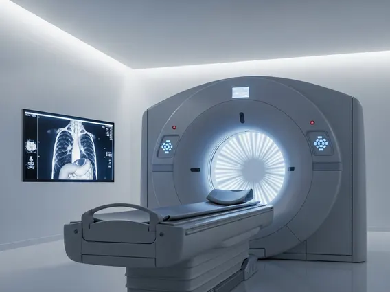
How does helical CT scan work? The process involves a patient lying on a motorized table that smoothly passes through a donut-shaped scanner called a gantry. Inside the gantry, an X-ray tube and a detector array rotate continuously around the patient. As the X-ray tube emits a fan-shaped beam, the detectors on the opposite side measure the X-rays that pass through the body. Simultaneously, the patient table moves, creating a continuous, spiral data acquisition path.
This continuous data stream is then sent to a powerful computer. The computer uses sophisticated algorithms to reconstruct the raw data into detailed cross-sectional images. This advanced processing is central to Helical CT imaging explanation, allowing for the creation of high-resolution two-dimensional images that can also be reprocessed into three-dimensional views of organs, bones, and blood vessels. The rapid acquisition and reconstruction capabilities allow clinicians to visualize complex anatomical structures with great clarity, aiding in the detection of diseases and injuries.
Uses and Benefits of Helical CT Scans
Helical CT scan uses and benefits are extensive, making it an indispensable tool in modern medicine. Its speed and ability to produce high-quality 3D images have made it a preferred method for diagnosing a wide range of conditions. For instance, in emergency medicine, helical CT can quickly identify internal injuries, fractures, and hemorrhages in trauma patients, enabling prompt treatment. In oncology, it is vital for detecting tumors, staging cancer, and monitoring treatment response.
The benefits of helical CT scans include:
- Reduced Scan Times: Faster acquisition minimizes patient discomfort and the risk of motion artifacts, particularly useful for pediatric or uncooperative patients.
- Improved Image Quality: Continuous data acquisition allows for better reconstruction of 3D images, providing more detailed anatomical information.
- Enhanced Detection: The spiral data path helps in detecting smaller lesions or abnormalities that might be missed with conventional, slice-by-slice scanning.
- Versatile Applications: Used across various specialties, including cardiovascular imaging for detecting blockages, neurological imaging for strokes, and musculoskeletal imaging for complex fractures.
- Dose Optimization: Modern helical CT scanners incorporate advanced dose modulation techniques to minimize radiation exposure while maintaining diagnostic image quality.
According to the World Health Organization (WHO), diagnostic imaging, including CT scans, plays a crucial role in improving global health outcomes by enabling early and accurate diagnosis of diseases, thereby facilitating timely and effective interventions.