Implant Displacement Views
Implant displacement views are specialized radiographic images crucial in medical diagnostics, particularly in orthopedics and other fields involving implanted devices. These views provide essential information regarding the position and stability of implants within the body.
Key Takeaways
- Implant Displacement Views are specialized X-ray techniques used to assess the precise position and stability of medical implants.
- Their primary clinical purpose is to detect complications such as migration, loosening, or malposition of implants.
- Proper interpretation involves comparing current views with baseline images and understanding anatomical landmarks.
- These views are vital for monitoring post-operative recovery and guiding potential revision surgeries.
- Accurate assessment requires a thorough understanding of imaging protocols and potential artifacts.
What is Implant Displacement Views and Its Clinical Purpose?
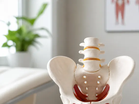
Implant Displacement Views refers to a specific set of radiographic imaging techniques designed to evaluate the precise location, orientation, and potential movement of medical implants within a patient’s body. These views are distinct from standard post-operative radiographs as they often involve specific patient positioning or dynamic imaging (e.g., flexion/extension views for spinal implants) to highlight potential displacement or instability that might not be apparent on static images. They are commonly utilized after procedures involving joint replacements (such as hip, knee, or shoulder), spinal instrumentation, dental implants, or other implanted devices where stability and correct positioning are critical for function and longevity.
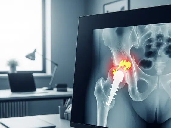
The Purpose of implant displacement views is multifaceted, primarily focusing on the early detection and characterization of implant-related complications. These complications can include implant migration, loosening, fracture, or impingement on surrounding anatomical structures, all of which can lead to pain, dysfunction, or implant failure. By providing detailed spatial information, these views help clinicians assess the integrity of the implant-bone interface, evaluate soft tissue relationships, and identify any subtle changes that might indicate a problem requiring intervention. For instance, in total hip arthroplasty, subtle changes in component position, particularly acetabular cup migration, can indicate early loosening, which, if left unaddressed, could lead to progressive pain and the need for complex revision surgery. Early detection of implant migration, as highlighted by studies, can significantly improve long-term outcomes and reduce the need for more invasive procedures.
Interpreting Implant Displacement Views: Key Considerations
Interpreting implant displacement views requires a systematic approach and a comprehensive understanding of both normal post-operative appearances and potential pathological findings. Clinicians typically compare current images with immediate post-operative baseline views and, if available, prior follow-up studies to identify any changes over time. Key considerations include assessing the alignment of the implant components relative to each other and to the surrounding bone, looking for evidence of lucency or sclerosis at the implant-bone interface, and evaluating for signs of component subsidence or migration.
Several factors are crucial when assessing these images. For example, in spinal fusion, assessing the stability of instrumentation involves looking for screw loosening, rod fracture, or cage subsidence. The presence of gas, fluid collections, or heterotopic ossification can also complicate interpretation and may indicate infection or other post-surgical issues. To aid in precise assessment, radiologists and orthopedic surgeons often look for specific indicators:
- Periprosthetic Lucency: A thin radiolucent line at the implant-bone interface, which can indicate loosening if it progresses or is wider than a certain threshold (e.g., >2mm).
- Component Migration: Any measurable change in the position of an implant component relative to fixed anatomical landmarks or previous images.
- Fracture: Evidence of fracture in the implant itself or in the surrounding bone (periprosthetic fracture).
- Impingement: Contact between implant components or between an implant and adjacent soft tissues or bone, which can cause pain or limited range of motion.
The detailed analysis provided by Implant displacement views explained helps guide clinical decisions, from conservative management to surgical revision. Accurate interpretation is paramount for patient safety and optimal long-term outcomes following implant surgery.