Core Needle Biopsy
A core needle biopsy is a common medical procedure used to diagnose various conditions, most notably cancer. It involves the removal of a small tissue sample for laboratory analysis, providing crucial information for diagnosis and treatment planning.

Key Takeaways
- Core Needle Biopsy is a minimally invasive procedure to extract tissue samples for diagnostic purposes.
- The procedure typically involves local anesthesia and imaging guidance to ensure precise tissue collection.
- Common risks are usually minor, such as bruising or discomfort, with serious complications being rare.
- Results are analyzed by a pathologist to determine if tissue is benign, malignant, or atypical.
- Understanding the results is crucial for guiding subsequent medical decisions and treatment plans.
What is Core Needle Biopsy?
Core Needle Biopsy refers to a diagnostic procedure where a small sample of tissue is removed from the body using a hollow needle. This technique is widely utilized to investigate suspicious lumps or abnormal areas identified through imaging tests, such as mammograms, ultrasounds, or CT scans. The primary purpose is to obtain tissue for microscopic examination by a pathologist, which helps determine the presence of disease, particularly cancer, or to characterize other conditions like infections or inflammatory processes. It is considered less invasive than a surgical biopsy, often performed on an outpatient basis, and provides more definitive results than fine needle aspiration, as it collects a larger tissue core.
The procedure is crucial for accurate diagnosis, allowing healthcare providers to differentiate between benign (non-cancerous) and malignant (cancerous) growths. For instance, according to the American Cancer Society, biopsies are essential for confirming cancer diagnoses, which then informs the most appropriate treatment strategy. By providing a clear tissue sample, a core needle biopsy helps avoid unnecessary surgery for benign conditions while ensuring timely intervention for malignant ones.
Core Needle Biopsy Procedure and Risks
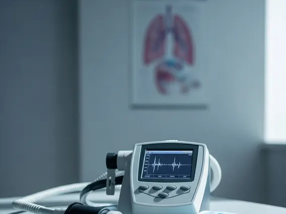
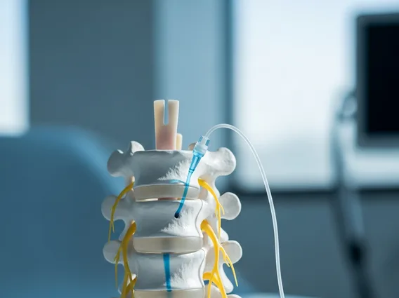
The core needle biopsy procedure typically begins with the patient lying comfortably, and the area to be biopsied is cleaned and numbed with a local anesthetic. Once the area is numb, a small incision may be made in the skin. The physician then uses a specialized hollow needle to extract several small tissue samples. To ensure accuracy, the needle’s insertion is often guided by imaging techniques such as ultrasound, mammography (for breast biopsies), or computed tomography (CT) scans, allowing the doctor to visualize the target area in real-time. After the samples are collected, the needle is removed, and pressure is applied to the site to minimize bleeding. A small dressing or bandage is then placed over the incision.
While generally safe, there are potential risks of core needle biopsy, though serious complications are uncommon. Patients may experience some discomfort, bruising, or minor bleeding at the biopsy site. Less frequently, risks can include:
- Infection, which can be managed with antibiotics.
- Excessive bleeding, particularly for individuals on blood-thinning medications.
- Damage to surrounding tissues or nerves, though this is rare due to imaging guidance.
- Pneumothorax (collapsed lung) in very rare cases of lung biopsies.
Patients are usually advised to avoid strenuous activity for a day or two following the procedure and to monitor the biopsy site for any signs of infection or excessive swelling.
Understanding Core Needle Biopsy Results
After the tissue samples are collected, they are sent to a pathology laboratory for analysis. Core needle biopsy results explained involve a pathologist examining the tissue under a microscope to identify any abnormal cells or structures. The time it takes to receive results can vary, typically ranging from a few days to over a week, depending on the complexity of the case and the tests required. Once the pathologist completes their analysis, they issue a report to the referring physician.
The results will generally fall into one of three categories:
- Benign: This indicates that no cancerous cells were found, and the growth or abnormality is non-cancerous.
- Malignant: This confirms the presence of cancerous cells, and the report will often specify the type and grade of cancer.
- Atypical or Indeterminate: These results suggest some abnormal cells are present, but they are not definitively cancerous. Further investigation, such as additional imaging or another biopsy, may be recommended.
It is crucial for patients to discuss their biopsy results thoroughly with their healthcare provider. The physician will explain what the results mean in the context of the patient’s overall health and recommend the next steps, which could range from continued monitoring to specific treatment plans. Understanding these results is a vital step in managing one’s health and making informed decisions about future care.