Coccyx
The Coccyx, commonly known as the tailbone, is a small, triangular bone located at the very bottom of the spine, below the sacrum. While often considered vestigial, it plays several important roles in human anatomy and biomechanics, providing support and serving as an attachment point for various structures.
Key Takeaways
- The Coccyx is the terminal segment of the vertebral column, composed of three to five fused vertebrae.
- It is located at the base of the spine, inferior to the sacrum, forming the posterior aspect of the pelvic floor.
- Despite its small size, the Coccyx provides crucial weight-bearing support, especially when sitting.
- It serves as an important attachment site for numerous muscles, ligaments, and tendons of the pelvic region.
- Understanding its anatomy and function is vital for diagnosing and treating conditions like coccydynia.
What is the Coccyx?
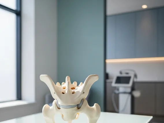
The Coccyx is a small, triangular bone that forms the very end of the spinal column in humans. Often referred to as the tailbone, it is typically composed of three to five separate or fused vertebral segments, though four is the most common number. These segments are usually fused by adulthood, creating a single, somewhat flexible bone. It is considered a remnant of a vestigial tail, but it has evolved to serve distinct functions in the human body.
The term Coccyx itself is derived from the Greek word “kokkyx,” meaning cuckoo, due to its perceived resemblance to a cuckoo’s beak. This bone plays a subtle yet significant role in supporting the body and facilitating various movements, particularly those involving the pelvic floor and lower limbs.
Coccyx Anatomy and Location
The coccyx anatomy and location are fundamental to understanding its function. Situated inferior to the sacrum, the Coccyx forms the most caudal (lowest) part of the vertebral column. It is nestled between the two halves of the pelvis, positioned just above the anal opening. Its triangular shape, with the base articulating with the sacrum and the apex pointing downwards, allows it to fit snugly within the pelvic girdle.
Anatomically, the Coccyx consists of several rudimentary vertebrae that progressively decrease in size from superior to inferior. The first coccygeal vertebra (Co1) is the largest and may remain unfused with the sacrum, forming the sacrococcygeal joint, which allows for limited movement. The subsequent vertebrae are typically fused. The Coccyx provides attachment for several important structures, including:
- Ligaments: Sacrococcygeal ligaments, anococcygeal ligament.
- Muscles: Gluteus maximus, coccygeus muscle, levator ani muscle.
- Tendons and fascia: Pelvic floor muscles and associated connective tissues.
Its position at the base of the spine makes it susceptible to injury, especially from direct trauma such as falls, which can lead to pain and discomfort known as coccydynia.
Function and Purpose of the Coccyx
The function of the coccyx extends beyond its vestigial origins, contributing significantly to human biomechanics and stability. One of its primary roles is to provide a weight-bearing tripod support when a person is sitting. Along with the ischial tuberosities (sit bones), the Coccyx helps to bear and distribute body weight, particularly when leaning back. This distribution helps to maintain balance and reduce pressure on other parts of the pelvis.
Furthermore, the coccyx meaning and purpose are closely tied to its role as an essential attachment point for various muscles and ligaments that form the pelvic floor. These muscles are crucial for supporting the pelvic organs, controlling defecation, and contributing to core stability. The Coccyx acts as an anchor for the following:
- The gluteus maximus, a large muscle responsible for hip extension and external rotation.
- The coccygeus muscle, which helps to support the pelvic floor.
- Parts of the levator ani muscle complex, vital for pelvic organ support and continence.
By serving as a stable point of origin and insertion for these structures, the Coccyx indirectly influences posture, movement, and the integrity of the pelvic region. While its range of motion is minimal, its structural presence is integral to the complex interplay of forces within the lower trunk.