
Diagnostic Mammogram
A diagnostic mammogram is a specialized X-ray examination of the breast used to investigate specific concerns or evaluate findings from a screening mammogram. Unlike routine screenings, it provides more detailed images to help diagnose potential breast conditions.
Key Takeaways
- A diagnostic mammogram is a targeted imaging test used to investigate specific breast symptoms or abnormal screening results.
- It differs from a screening mammogram, which is a routine check for asymptomatic individuals.
- The procedure involves more detailed views and often includes magnification or spot compression.
- Results are frequently discussed with the patient immediately after the examination.
- Early and accurate diagnosis through this method is crucial for effective treatment planning.
What is a Diagnostic Mammogram?
A diagnostic mammogram is an advanced imaging procedure performed when a woman has a specific breast concern or when an abnormality is detected during a routine screening mammogram. This type of mammogram focuses on a particular area of interest, providing radiologists with more detailed images to assess the nature of a breast change. Its primary purpose of a diagnostic mammogram is to determine if a suspicious area is benign (non-cancerous) or malignant (cancerous).
Common reasons for undergoing a diagnostic mammogram include evaluating a new lump or mass, persistent breast pain, nipple discharge, skin changes on the breast, or an area of concern identified on a prior screening mammogram. The enhanced imaging capabilities allow for a thorough examination, often involving additional views and techniques not typically used in screening.
Diagnostic vs. Screening Mammograms
Understanding the distinction between a diagnostic mammogram and a screening mammogram is crucial. While both use X-rays to create images of the breast, their objectives and procedures differ significantly. A screening mammogram vs screening mammogram comparison highlights these differences in their application and intent.
A screening mammogram is a routine preventive measure performed on women who have no symptoms or signs of breast cancer. Its goal is to detect breast cancer early, often before a lump can be felt, when it is most treatable. According to the Centers for Disease Control and Prevention (CDC), regular screening mammograms are recommended for women aged 40 and older, though specific guidelines may vary based on individual risk factors.
In contrast, a diagnostic mammogram is performed when there is a specific reason to investigate. It is not a routine check but a targeted examination. The table below summarizes the key differences:
| Feature | Screening Mammogram | Diagnostic Mammogram |
|---|---|---|
| Purpose | Routine check for early detection in asymptomatic women | Investigate specific symptoms or abnormal screening findings |
| Symptoms | No symptoms present | Presence of symptoms (lump, pain, discharge) or abnormal finding |
| Views | Standard views (usually two per breast) | Additional, targeted, and magnified views as needed |
| Radiologist Presence | Typically reviewed later by a radiologist | Often reviewed by a radiologist during the exam for immediate guidance |
| Duration | Shorter (approx. 15-20 minutes) | Longer (approx. 30-60 minutes or more) |
The Diagnostic Mammogram Procedure
When undergoing a diagnostic mammogram, patients can expect a more in-depth examination compared to a standard screening. The procedure is designed to provide comprehensive images of the area of concern. Before the exam, it’s important to inform the technologist about any symptoms you are experiencing or the specific area that needs evaluation.
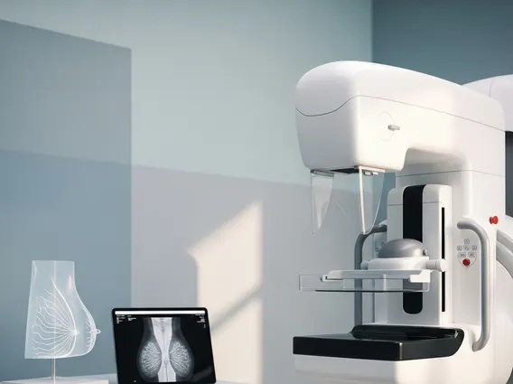
During the procedure, you will be asked to position your breast on a platform, and compression will be applied. While this can be uncomfortable, it is essential for obtaining clear images and reducing radiation dose. Unlike screening mammograms, which typically take two views of each breast, a diagnostic mammogram may involve multiple additional views, including:
- Spot Compression Views: These use a smaller compression paddle to apply more pressure to a specific area, spreading out breast tissue for better visualization.
- Magnification Views: These provide a magnified image of a small area, helping to assess microcalcifications (tiny calcium deposits) or subtle masses.
- Latero-Medial or Medio-Lateral Oblique Views: These are additional angles to better visualize the area of concern.
The technologist works closely with a radiologist, who may be present during the exam to guide the imaging process in real-time. This immediate oversight allows for adjustments to be made, ensuring all necessary images are captured. After the images are taken, the radiologist will review them, and in many cases, preliminary results can be discussed with you on the same day. This immediate feedback helps alleviate anxiety and allows for prompt next steps if further evaluation, such as an ultrasound or biopsy, is recommended.



















