
Digital Mammography
Digital Mammography is an advanced imaging technique used for breast cancer screening and diagnosis. It offers several advantages over traditional film mammography, providing clearer images and improved detection capabilities.
Key Takeaways
- Digital Mammography uses X-rays and computer technology to create detailed images of breast tissue.
- It is a crucial tool for early detection of breast cancer, often before a lump can be felt.
- The process involves compressing the breast and taking X-ray images, which are then stored and analyzed digitally.
- Key benefits include improved image quality, reduced radiation dose, and enhanced diagnostic accuracy, especially for certain breast types.
- Digital mammography offers significant advantages compared to traditional film-based methods, particularly in image manipulation and storage.
What is Digital Mammography?
Digital Mammography refers to a specialized type of mammography that uses digital receptors and computers instead of X-ray film to examine breast tissue. This technology converts X-ray energy into digital images, which can then be viewed on a computer monitor, manipulated, and stored electronically. It is a primary screening tool for breast cancer, recommended for women typically starting at age 40, though guidelines can vary by country and individual risk factors. The American Cancer Society recommends annual mammograms for women at average risk beginning at age 40 and continuing as long as they are in good health. This advanced imaging technique plays a vital role in the early detection of breast abnormalities, which is crucial for successful treatment outcomes.
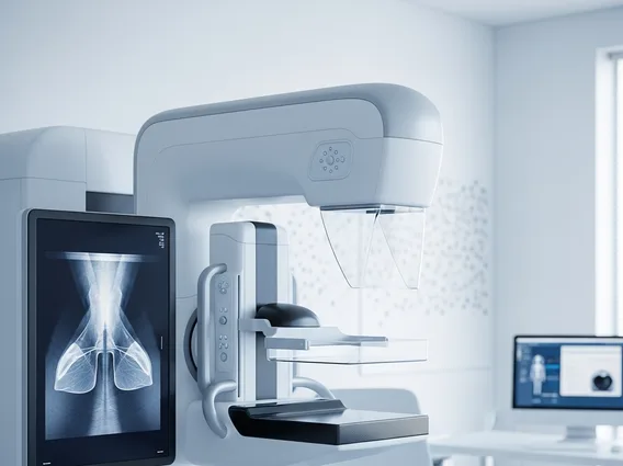
How Digital Mammography Works
The process of how digital mammography works involves several key steps designed to capture high-quality images of the breast. During the procedure, the patient’s breast is positioned on a special platform and gently compressed using a clear plastic paddle. This compression is essential for several reasons: it flattens the breast tissue to ensure all areas are visible, reduces the amount of radiation needed, and minimizes motion blur. An X-ray tube then generates a low dose of radiation that passes through the breast tissue. Instead of exposing film, a digital detector captures the X-ray signals and converts them into electrical signals. These signals are then processed by a computer to create detailed digital images, which are immediately available for review by a radiologist. The ability to adjust contrast, zoom in on specific areas, and invert colors digitally significantly enhances the radiologist’s ability to detect subtle changes or abnormalities.
Benefits and Comparison to Traditional Mammography
The benefits of digital mammography are numerous, leading to its widespread adoption over traditional film-based methods. One significant advantage is superior image quality, which allows radiologists to more easily detect subtle lesions and microcalcifications, particularly in dense breast tissue. Digital images can be manipulated post-acquisition, meaning brightness, contrast, and zoom can be adjusted without re-exposing the patient to radiation. This flexibility can reduce the need for repeat mammograms. Furthermore, digital mammography typically involves a slightly lower radiation dose compared to traditional film mammography, enhancing patient safety.
When considering digital mammography vs traditional film mammography, several distinctions become apparent:
- Image Acquisition: Digital systems use electronic detectors, while traditional systems use X-ray film.
- Image Quality: Digital images often provide better contrast and clarity, especially for women with dense breasts or those who are premenopausal.
- Radiation Dose: Digital mammography generally uses a lower radiation dose.
- Storage and Sharing: Digital images are easily stored, retrieved, and shared electronically, facilitating consultations and reducing physical storage needs.
- Workflow Efficiency: Digital systems offer faster image acquisition and review, improving clinic workflow.
| Feature | Digital Mammography | Traditional Mammography |
|---|---|---|
| Image Medium | Digital detectors, computer files | X-ray film |
| Image Manipulation | Adjustable brightness, contrast, zoom post-acquisition | Fixed image, no post-acquisition adjustment |
| Radiation Dose | Generally lower | Slightly higher |
| Image Storage | Electronic, easily archived and shared | Physical film, requires dedicated storage |
| Detection in Dense Breasts | Improved | Challenging |
This comparison underscores why digital mammography has become the standard for breast cancer screening, offering enhanced diagnostic capabilities and operational efficiencies.


