Digital Photography
Digital Photography, in a clinical context, refers to the application of digital imaging technology for diagnostic, monitoring, and documentation purposes within healthcare settings. This method offers a precise and efficient way to capture visual information relevant to patient care.
Key Takeaways
- Digital Photography is a vital clinical tool for documenting patient conditions, tracking progress, and aiding diagnosis.
- Digital cameras for medical use capture light through a lens onto a sensor, converting it into digital data for high-resolution image storage.
- Proper lighting, focus, and consistent framing are crucial for obtaining accurate and reproducible clinical images.
- Adhering to patient consent and data privacy protocols is paramount when utilizing digital photography in healthcare.
- Effective image management and secure storage are essential for maintaining patient records and facilitating interdisciplinary communication.
What is Digital Photography?
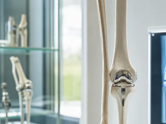
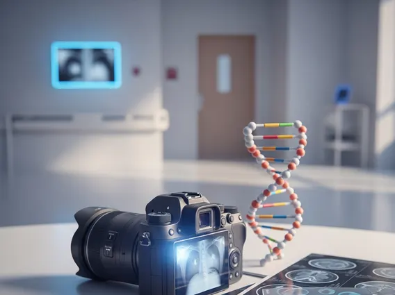
Digital Photography is a specialized application of digital imaging technology within the medical field, primarily used for capturing, storing, and transmitting high-resolution images of patients, anatomical structures, or clinical procedures. This technique is invaluable for documenting various conditions, such as dermatological lesions, wound healing, surgical sites, and dental anomalies, providing a visual record that complements written medical notes. Its utility extends to patient education, research, and interdisciplinary consultations, enhancing communication among healthcare professionals.
The transition from traditional film to digital methods has significantly improved efficiency and accessibility in clinical documentation. Digital images can be instantly reviewed, easily shared securely, and integrated into electronic health records (EHRs), facilitating longitudinal tracking of patient progress. This capability supports more accurate diagnoses and personalized treatment plans, making Digital Photography an indispensable tool in modern medicine.
How Digital Cameras Work
Digital cameras, when employed in a medical setting, operate on fundamental principles to convert light into digital data. At their core, these devices utilize a lens system to focus light from the subject onto an image sensor, typically a Charge-Coupled Device (CCD) or Complementary Metal-Oxide-Semiconductor (CMOS). Each pixel on the sensor converts light into an electrical charge, which is then processed by an analog-to-digital converter (ADC) to create a digital image file.
Key components for clinical use include high-resolution sensors, which ensure detailed capture of subtle anatomical features, and specialized lenses designed for macro photography or specific medical applications. Advanced image processors within the camera optimize color accuracy and exposure, crucial for consistent and reliable documentation. The resulting digital files, often in formats like JPEG or TIFF, are then stored on memory cards or transferred directly to secure hospital networks, ready for review and integration into patient records.
Getting Started: Digital Photography Tips for Beginners
For healthcare professionals new to using digital photography in a clinical context, a structured approach is essential to ensure high-quality, ethically sound, and medically useful images. This section serves as a beginner’s guide to digital photography for clinical applications, focusing on practical considerations.
Before any image capture, obtaining informed patient consent is paramount, clearly explaining the purpose and storage of the photographs. Proper lighting is critical; natural, diffused light is often ideal, but consistent artificial lighting setups can minimize shadows and ensure accurate color representation. Maintaining a consistent distance and angle from the subject for follow-up images allows for reliable comparison over time. Here are some essential digital photography tips for beginners:
- Patient Consent: Always secure explicit, informed consent from the patient before taking any photographs, ensuring they understand the purpose and use of the images.
- Consistent Framing: For serial documentation, strive for identical framing, distance, and angle in every subsequent photograph to allow for accurate comparison of changes.
- Optimal Lighting: Utilize consistent, shadow-free lighting. Ring flashes or twin flashes are often used in dermatology and dentistry to provide even illumination.
- Sharp Focus: Ensure images are sharply focused on the area of interest. Many modern cameras offer autofocus systems that can assist, but manual focus may be necessary for very specific details.
- White Balance: Set the camera’s white balance correctly for the lighting conditions to ensure accurate color rendition, which is vital for assessing skin tones or lesion characteristics.
- Secure Storage: Implement robust data management protocols for secure storage and retrieval of images, adhering to all privacy regulations (e.g., HIPAA in the U.S.).
Mastering these techniques ensures that digital images contribute effectively to patient care and medical documentation.