Dxa
Dual-energy X-ray Absorptiometry, commonly known as DXA, is a crucial diagnostic tool in modern medicine. It provides precise measurements of bone mineral density, playing a vital role in assessing bone health and detecting conditions like osteoporosis.
Key Takeaways
- DXA is a non-invasive imaging test that measures bone mineral density.
- It is the gold standard for diagnosing osteoporosis and assessing fracture risk.
- The procedure is quick, painless, and involves minimal radiation exposure.
- DXA helps monitor bone health over time and evaluate treatment effectiveness.
- Early detection through DXA can significantly improve patient outcomes.
What is DXA (Dual-energy X-ray Absorptiometry)?
DXA, or Dual-energy X-ray Absorptiometry, is a sophisticated imaging technology used primarily to measure bone mineral density (BMD). It is considered the most accurate and widely used method for diagnosing osteoporosis and assessing an individual’s risk of developing fractures. A DXA scan utilizes two distinct X-ray beams with different energy levels to differentiate between bone and soft tissue, allowing for a precise measurement of bone density in specific areas, most commonly the hip, spine, and forearm. The information gathered from a DXA scan helps healthcare providers understand the strength and health of a patient’s bones.
The principle behind what is Dxa involves passing a very low-dose X-ray through the body. The amount of X-ray energy absorbed by the bone is then measured by a detector. Denser bones absorb more X-rays, while less dense bones absorb less. This data is then processed by a computer to generate images and numerical scores (T-score and Z-score) that indicate the bone mineral density. These scores are compared to those of healthy young adults (T-score) or age-matched individuals (Z-score) to determine if bone density is normal, low (osteopenia), or significantly low (osteoporosis).
The DXA Scan Procedure: What to Expect
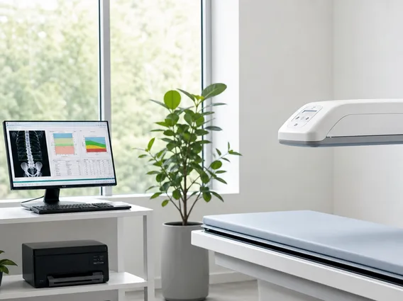
The DXA scan procedure is a straightforward, non-invasive, and typically quick outpatient test. Patients generally do not need to make extensive preparations, though they may be advised to avoid calcium supplements for 24 hours prior to the test. Upon arrival, the patient will lie comfortably on a padded table. A scanner arm will then pass over the body, emitting low-dose X-rays. The entire process usually takes between 10 to 20 minutes, depending on the areas being scanned. During the scan, patients are asked to remain still to ensure clear and accurate images.
To ensure the DXA bone density test explained clearly, here are the general steps involved:
- Preparation: Patients wear loose, comfortable clothing and remove any metal objects (jewelry, zippers, buttons) that could interfere with the X-ray images.
- Positioning: The technologist will carefully position the patient on the scanning table, often using foam blocks to help maintain the correct posture for accurate measurements of the hip and spine.
- Scanning: The scanner arm moves slowly over the body, capturing images. The radiation exposure is minimal, significantly less than a standard chest X-ray.
- Results: After the scan, the images and data are analyzed by a radiologist or other qualified physician, who then provides a report to the referring doctor.
Patients typically experience no pain or discomfort during the procedure and can resume their normal activities immediately afterward.
Uses and Benefits of DXA Bone Density Testing
The primary DXA scan uses and benefits revolve around the early detection, diagnosis, and monitoring of bone-related conditions. It is the gold standard for diagnosing osteoporosis, a condition characterized by brittle bones that are prone to fractures. According to the International Osteoporosis Foundation, osteoporosis causes more than 8.9 million fractures annually worldwide, meaning an osteoporotic fracture occurs every 3 seconds. Early diagnosis through DXA allows for timely intervention, which can significantly reduce the risk of debilitating fractures.
Key uses and benefits include:
- Diagnosing Osteoporosis and Osteopenia: DXA accurately identifies reduced bone density before a fracture occurs.
- Assessing Fracture Risk: The test helps predict an individual’s likelihood of future fractures based on their bone density.
- Monitoring Treatment Effectiveness: For patients undergoing treatment for osteoporosis, regular DXA scans can track changes in bone density and assess how well the treatment is working.
- Identifying High-Risk Individuals: It is particularly recommended for postmenopausal women, men over 70, and individuals with certain medical conditions or taking medications that affect bone health.
- Guiding Lifestyle Recommendations: Results can inform dietary and exercise recommendations to improve bone health.
By providing precise and reliable measurements, DXA bone density testing empowers both patients and healthcare providers to make informed decisions regarding bone health management, ultimately contributing to a better quality of life and reduced fracture incidence.