Plexiform Fibrohistiocytic Tumor
Plexiform Fibrohistiocytic Tumor is a rare, low-grade malignant soft tissue tumor that typically affects children and young adults. Understanding this condition is crucial for timely diagnosis and effective management.

Key Takeaways
- Plexiform Fibrohistiocytic Tumor is a rare, low-grade malignant soft tissue tumor primarily affecting young individuals.
- It commonly presents as a slow-growing, firm nodule, often on the extremities.
- Diagnosis relies on biopsy and histopathological examination, supported by imaging studies.
- The primary treatment is surgical removal, with recurrence being a possibility.
- Prognosis is generally favorable, though long-term follow-up is essential due to potential for local recurrence.
What is Plexiform Fibrohistiocytic Tumor?
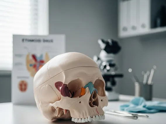
Plexiform Fibrohistiocytic Tumor is a distinctive, rare soft tissue neoplasm characterized by its intermediate malignant potential. It primarily affects the dermis and subcutaneous tissues, most commonly found on the extremities, head, and neck. This tumor is predominantly observed in children and young adults, with a slight female predominance. Its unique histological features include a plexiform (net-like) growth pattern of spindle cells and histiocyte-like cells, often accompanied by osteoclast-like giant cells. The World Health Organization (WHO) classifies it as a tumor of intermediate malignancy, meaning it has a potential for local recurrence and, very rarely, distant metastasis.
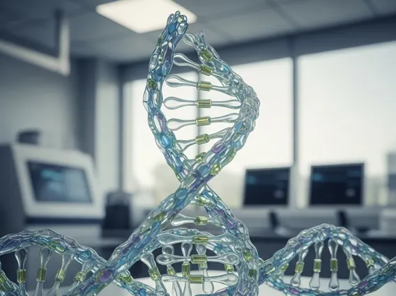
The exact cause of Plexiform Fibrohistiocytic Tumor remains unknown, but genetic studies have identified specific chromosomal translocations, such as t(1;10)(p31;q24), in a subset of cases, suggesting a molecular basis for its development. This understanding helps in confirming diagnosis and distinguishing it from other soft tissue tumors. Despite its malignant classification, it generally exhibits a less aggressive clinical course compared to high-grade sarcomas, making accurate diagnosis vital for appropriate patient management.
Symptoms and Diagnosis of Plexiform Fibrohistiocytic Tumor
The presentation of Plexiform Fibrohistiocytic Tumor is typically insidious, with patients often noticing a slowly enlarging, firm, and sometimes painless nodule. While pain or tenderness can occur, it is less common. The tumor can range in size from a few millimeters to several centimeters and is usually mobile, though it can become fixed to deeper structures in advanced cases. The most common sites are the upper and lower extremities, but it can appear anywhere on the body.
The process for plexiform fibrohistiocytic tumor symptoms diagnosis involves a combination of clinical evaluation, imaging studies, and definitive histopathological examination. Initial assessment often includes:
- Clinical Examination: Palpation of the mass to assess size, consistency, mobility, and any associated pain.
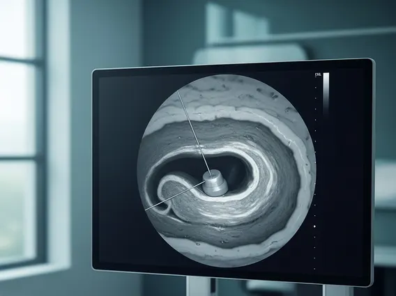
- Imaging Studies: Ultrasound can determine the solid nature and depth of the lesion. Magnetic Resonance Imaging (MRI) is often used to define the tumor’s exact size, extent, and relationship to surrounding tissues, which is crucial for surgical planning.
- Biopsy: A definitive diagnosis requires a tissue biopsy, which can be incisional (a portion of the tumor) or excisional (removal of the entire tumor). The biopsy specimen undergoes detailed histopathological analysis by a pathologist.
- Immunohistochemistry: Special stains are used to identify specific protein markers within the tumor cells, aiding in differentiation from other soft tissue tumors.
- Genetic Analysis: In some cases, molecular testing for specific genetic translocations can confirm the diagnosis, especially in challenging presentations.
Accurate diagnosis is paramount to distinguish Plexiform Fibrohistiocytic Tumor from other benign or more aggressive malignant soft tissue lesions, ensuring that patients receive the most appropriate and effective treatment plan.
Treatment and Prognosis for Plexiform Fibrohistiocytic Tumor
The primary and most effective approach for plexiform fibrohistiocytic tumor treatment options is complete surgical excision. This typically involves wide local excision, where the tumor is removed along with a margin of healthy surrounding tissue to minimize the risk of local recurrence. The goal is to achieve clear surgical margins, which significantly improves outcomes. In cases where complete excision is challenging due to the tumor’s location or size, a staged approach or more extensive surgery may be necessary.
Adjuvant therapies, such as chemotherapy or radiation therapy, are generally not indicated for Plexiform Fibrohistiocytic Tumor due to its low-grade nature and limited metastatic potential. These therapies are usually reserved for very rare instances of aggressive behavior, incomplete excision with positive margins where further surgery is not feasible, or documented distant metastasis. However, such scenarios are exceedingly rare. Regular follow-up after surgery is crucial to monitor for any signs of local recurrence, which can occur in a significant percentage of cases, ranging from 20% to 50% according to various studies, often years after initial treatment. This emphasizes the need for long-term surveillance.
The overall plexiform fibrohistiocytic tumor prognosis is generally favorable, given its low metastatic rate and high survival rates. Most patients achieve a good outcome with appropriate surgical management. However, the potential for local recurrence necessitates diligent follow-up, typically involving regular clinical examinations and imaging of the surgical site. While distant metastases are exceptionally rare, occurring in less than 5% of cases, they can impact the prognosis. Patients are usually advised to maintain regular contact with their oncology team for several years post-treatment to ensure early detection and management of any recurrent disease, thereby optimizing long-term health outcomes.