Indirect Ophthalmoscopy
Indirect Ophthalmoscopy is a crucial diagnostic procedure used in ophthalmology to examine the health of the retina, optic disc, macula, and vitreous humor. This comprehensive examination allows eye care professionals to detect and monitor various eye conditions affecting the posterior segment of the eye.
Key Takeaways
- Indirect Ophthalmoscopy is a comprehensive eye exam technique for viewing the back of the eye.
- It uses a bright light source and a condensing lens to provide a wide, stereoscopic view of the retina.
- The procedure is vital for diagnosing conditions like retinal detachments, glaucoma, and diabetic retinopathy.
- Patients typically experience temporary blurred vision and light sensitivity due to pupil dilation.
- While generally safe, minor risks include temporary discomfort and light sensitivity.
What is Indirect Ophthalmoscopy?
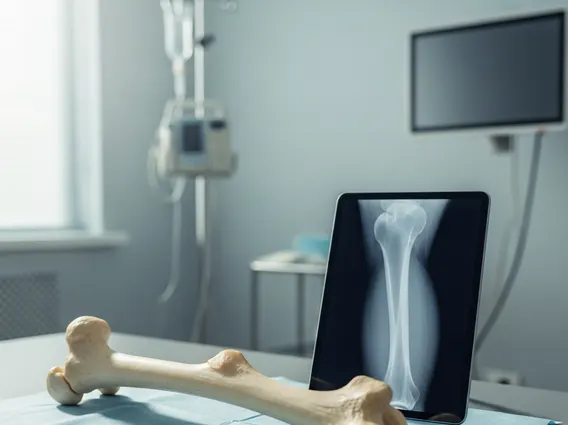
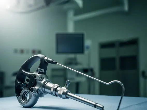
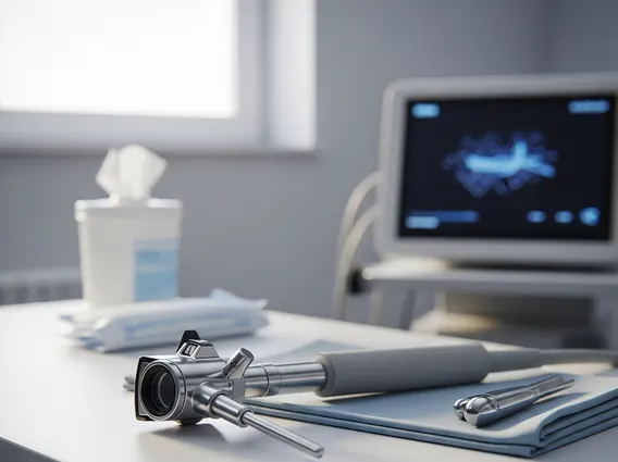
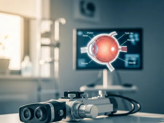
Indirect Ophthalmoscopy refers to a specialized eye examination technique that allows an ophthalmologist or optometrist to view the retina and other structures at the back of the eye in great detail. Unlike direct ophthalmoscopy, which provides a magnified, upright but smaller view, indirect ophthalmoscopy uses a bright light source worn on the examiner’s head and a handheld condensing lens to create an inverted, magnified, and wider field of view. This stereoscopic (3D) perspective is invaluable for assessing the entire retina, including its peripheral areas, which are often difficult to visualize with other methods.
Purpose and Procedure of Indirect Ophthalmoscopy
The purpose of indirect ophthalmoscopy exam is to thoroughly evaluate the posterior segment of the eye, aiding in the diagnosis and management of numerous ocular and systemic conditions. It is particularly effective for identifying retinal detachments, tears, holes, diabetic retinopathy, macular degeneration, glaucoma, and tumors. This examination is often a standard part of a comprehensive eye check-up, especially for individuals at higher risk for certain eye diseases.
The indirect ophthalmoscopy procedure details typically involve several steps to ensure a clear and comprehensive view. First, the patient’s pupils are usually dilated using eye drops. This widening of the pupils allows the examiner to see more of the retina. Once the pupils are dilated, the patient sits comfortably, and the examiner positions themselves in front of them. The examiner wears a head-mounted ophthalmoscope, which projects a bright light into the patient’s eye. While holding a condensing lens a few inches from the eye, the examiner systematically scans the retina. Patients are often asked to look in various directions to allow the examiner to visualize different parts of the retina. The entire procedure usually takes about 5-10 minutes per eye, though the effects of dilation can last for several hours, causing light sensitivity and blurred near vision.
Benefits and Potential Risks of Indirect Ophthalmoscopy
The indirect ophthalmoscopy benefits and risks make it a preferred method for many diagnostic scenarios. One of the primary benefits is the ability to obtain a wide, stereoscopic view of the retina, which is crucial for detecting subtle changes or abnormalities, especially in the peripheral retina. This broad view significantly enhances the early detection of conditions like retinal tears or detachments, which can be sight-threatening if not addressed promptly. It also allows for a better assessment of the optic nerve head and macula, vital structures for vision. The procedure is non-invasive and generally well-tolerated by patients.
While generally safe, there are some potential risks and side effects associated with indirect ophthalmoscopy, primarily due to the pupil dilation. These include:
- Temporary blurred vision: Near vision is particularly affected, making reading or close work difficult for several hours.
- Increased light sensitivity (photophobia): Pupils remain dilated, allowing more light into the eyes, which can be uncomfortable in bright environments.
- Rare allergic reaction: To the dilating eye drops, though this is uncommon.
- Acute angle-closure glaucoma: An extremely rare complication in individuals with narrow anterior chamber angles, where pupil dilation can trigger a sudden increase in eye pressure.
Patients are typically advised not to drive immediately after the procedure and to wear sunglasses to mitigate light sensitivity until the effects of the dilation drops wear off.