Image Guided Radiation Therapy
Image Guided Radiation Therapy (IGRT) represents a significant advancement in cancer treatment, enhancing the precision and effectiveness of radiation delivery. This sophisticated technique allows oncologists to target tumors with greater accuracy while minimizing damage to surrounding healthy tissues.
Key Takeaways
- Image Guided Radiation Therapy (IGRT) precisely targets tumors using advanced imaging during radiation treatment.
- It enables real-time adjustments to radiation beams, accounting for tumor movement and anatomical changes.
- This enhanced precision helps deliver optimal radiation doses to cancer cells while safeguarding surrounding healthy organs.
- IGRT can lead to improved treatment outcomes and a reduction in radiation-related side effects.
What is Image Guided Radiation Therapy (IGRT)?
Image Guided Radiation Therapy (IGRT) is a sophisticated radiation treatment technique that uses frequent imaging during a course of radiation therapy to improve the precision and accuracy of treatment delivery. The primary goal of IGRT is to ensure that radiation beams are directed precisely at the tumor while sparing healthy surrounding tissues and organs. This approach is crucial because tumors can move between treatments or even during a single treatment session due to physiological processes like breathing, digestion, or changes in organ filling.
The continuous monitoring and adjustment capabilities offered by IGRT allow clinicians to adapt the treatment plan in real-time. This dynamic approach ensures that the radiation dose is consistently delivered to the intended target, even if the tumor’s position shifts. An effective image guided radiation therapy explanation emphasizes its role in enhancing dose conformity, meaning the radiation dose closely matches the shape of the tumor, thereby maximizing tumor control and minimizing toxicity to normal tissues.
How Does IGRT Work?
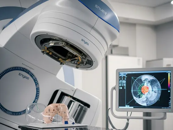
IGRT integrates advanced imaging technologies directly into the radiation treatment process. Before or during each treatment fraction, imaging scans are performed to visualize the tumor and surrounding anatomy. These images are then compared to the initial planning scans, allowing the radiation oncology team to detect any shifts in the tumor’s position or changes in the patient’s internal anatomy. Based on these comparisons, precise adjustments can be made to the patient’s position on the treatment couch or to the radiation beam’s trajectory.
The imaging modalities used in IGRT vary, offering different levels of detail and real-time capabilities. Common imaging techniques include:
- Cone-beam Computed Tomography (CBCT): Provides 3D images of the patient’s anatomy, allowing for precise alignment before each treatment.
- Kilovoltage (kV) X-rays: Offer high-resolution 2D images, often used for daily verification of bone structures and fiducial markers.
- Megavoltage (MV) X-rays: Can be used for imaging through the treatment beam, providing verification of the treatment field.
- Ultrasound: Useful for visualizing soft tissues, particularly in the prostate, without additional radiation exposure.
- Magnetic Resonance Imaging (MRI): Increasingly integrated into IGRT systems, offering superior soft-tissue contrast for precise tumor tracking.
By using these imaging tools, IGRT systems can account for organ motion, such as lung tumors moving with respiration, or changes in bladder and rectal filling, ensuring that the radiation dose is always delivered accurately to the target volume.
Benefits of Image Guided Radiation Therapy
The primary benefits of Image Guided Radiation Therapy stem from its ability to deliver radiation with unprecedented precision. This enhanced accuracy allows for higher, more effective doses of radiation to be delivered directly to the tumor, increasing the likelihood of tumor control and eradication. Concurrently, the ability to spare more healthy tissue means a significant reduction in treatment-related side effects, improving the patient’s quality of life both during and after therapy. For instance, in prostate cancer treatment, IGRT can reduce the risk of rectal and bladder toxicity by precisely targeting the prostate gland while avoiding adjacent organs.
Furthermore, IGRT enables the treatment of tumors located in challenging areas, such as those near critical organs or in regions prone to significant movement. This technology is particularly valuable for hypofractionated radiation therapy, where fewer, larger doses are delivered, making precision even more critical. According to the American Society for Radiation Oncology (ASTRO), the adoption of advanced techniques like IGRT has significantly contributed to the improved safety and efficacy of modern radiation therapy. This continuous refinement in treatment delivery underscores IGRT’s role in advancing oncology care, offering patients more targeted and less toxic treatment options.