
Burr Hole
A Burr Hole is a neurosurgical procedure involving the creation of a small opening in the skull. This common technique allows surgeons access to the brain for various diagnostic and therapeutic purposes, often serving as a critical intervention in neurological emergencies.
Key Takeaways
- A Burr Hole is a small opening drilled into the skull to access the brain.
- Its primary purpose is to relieve pressure, drain fluid, or facilitate further surgical interventions.
- The procedure is typically performed under general anesthesia, involving careful preparation and precise execution.
- Common applications include treating subdural hematomas and hydrocephalus.
- Recovery involves monitoring for complications and managing post-operative symptoms.
What is a Burr Hole Procedure and Its Purpose?
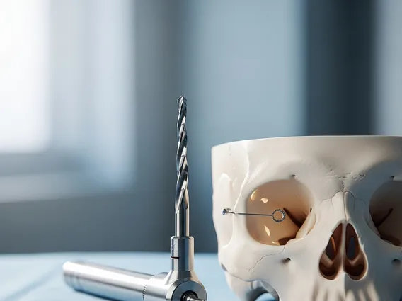
A Burr Hole procedure involves drilling a small hole, typically 10-14 mm in diameter, through the skull bone. This neurosurgical technique is fundamental for accessing the intracranial space, allowing surgeons to address various conditions affecting the brain and its surrounding structures. It is a minimally invasive approach compared to larger craniotomies, offering a direct pathway to the brain while minimizing trauma to surrounding tissues.
The purpose of burr hole in brain surgery is multifaceted, primarily focusing on relieving pressure, draining accumulated fluids, or providing a port for more complex procedures. One of its most common applications is in treating subdural hematomas, where blood collects between the brain and its outermost covering (dura mater), causing dangerous pressure on the brain. By creating a burr hole, surgeons can insert a catheter to drain the blood, thereby alleviating intracranial pressure and preventing further neurological damage.
Other critical applications include:
- Drainage of Abscesses: Removing pus from brain infections.
- Placement of Intracranial Pressure (ICP) Monitors: Inserting devices to continuously measure pressure inside the skull, crucial for managing severe head injuries or conditions like hydrocephalus.
- Biopsy: Obtaining tissue samples from brain lesions for diagnostic analysis.
- Ventriculostomy: Creating an opening into the brain’s ventricles to drain cerebrospinal fluid (CSF) in cases of hydrocephalus or to relieve acute hydrocephalus.
This procedure is often a life-saving intervention, rapidly addressing conditions that could otherwise lead to severe neurological impairment or death.
The Burr Hole Surgical Process
The burr hole surgery explanation details a precise surgical process, typically performed under general anesthesia, though local anesthesia with sedation may be used in some cases. Before the procedure, the patient undergoes thorough medical evaluation, including imaging studies like CT scans or MRI, to pinpoint the exact location for the burr hole. This meticulous planning ensures accuracy and minimizes risks.
The process begins with the patient positioned appropriately, and the surgical site on the scalp is meticulously cleaned and sterilized. The surgeon then injects a local anesthetic into the scalp, even if general anesthesia is used, to further reduce pain and bleeding. An incision is made through the scalp, exposing the skull bone. Using a specialized surgical drill, a small, circular opening is carefully made through the bone, penetrating the outer layer of the skull without damaging the underlying dura mater.
Once the burr hole is created, the dura mater may be carefully incised to access the subdural or subarachnoid space, or the brain tissue itself, depending on the surgical objective. For instance, in cases of subdural hematoma, a small catheter is inserted through the burr hole to drain the accumulated blood. After the necessary intervention is completed, the catheter may be left in place temporarily, or the burr hole may be covered with a small plate or left to heal naturally. The scalp incision is then closed with sutures or staples. Patients are closely monitored in the post-operative period for any signs of complications, such as infection, bleeding, or changes in neurological status.
This approach allows for targeted intervention with minimal disruption to surrounding healthy brain tissue, facilitating quicker recovery times compared to more extensive cranial surgeries.


