
Stage Iiic Intraocular Melanoma Of The Ciliary Body And Choroid
Stage IIIC Intraocular Melanoma of the Ciliary Body and Choroid represents an advanced form of a rare eye cancer, specifically affecting critical structures within the eye responsible for vision and eye health. Understanding this condition is crucial for effective management and improved patient outcomes.
Key Takeaways
- Stage IIIC Intraocular Melanoma of the Ciliary Body and Choroid is an advanced, rare eye cancer affecting the uvea.
- Symptoms can be subtle, including vision changes, and diagnosis relies on comprehensive ophthalmological exams and imaging.
- Treatment often involves radiation therapy or surgery, aiming to preserve vision and prevent spread.
- Prognosis is influenced by tumor characteristics, with early detection and aggressive treatment being vital.
- Regular follow-up is essential due to the risk of recurrence and metastasis.
What is Stage IIIC Intraocular Melanoma of the Ciliary Body and Choroid?
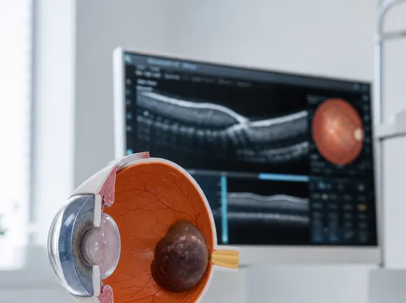
Stage IIIC Intraocular Melanoma of the Ciliary Body and Choroid refers to a specific classification of a malignant tumor originating in the uvea, the middle layer of the eye. The uvea comprises the iris, ciliary body, and choroid. In this advanced stage, the melanoma has grown significantly within the ciliary body and choroid, which are crucial for producing aqueous humor and supplying blood to the retina, respectively. The “IIIC” designation typically indicates a large tumor size with specific features, such as extraocular extension (spread outside the eyeball) or involvement of regional lymph nodes, but without distant metastasis at the time of diagnosis.
This condition is a rare form of cancer, making it the most common primary intraocular malignancy in adults, yet its overall incidence is low. According to the American Cancer Society, approximately 2,000 adults are diagnosed with ocular melanoma in the United States each year. The ciliary body and choroid are common sites for these melanomas due to their rich blood supply and melanocyte presence. Understanding the specific staging, such as Stage IIIC, is vital as it guides treatment strategies and provides insight into the potential prognosis.
Symptoms and Diagnosis of Stage IIIC Intraocular Melanoma
The Stage IIIC intraocular melanoma symptoms can be subtle, especially in earlier stages, making regular comprehensive eye examinations critical. As the tumor progresses to Stage IIIC, symptoms may become more noticeable due to its larger size and potential impact on ocular structures. Common symptoms can include:
- Blurred vision or decreased visual acuity
- Visual field defects, such as a blind spot
- Flashes of light (photopsia) or floaters
- Changes in the shape of the pupil
- Eye pain, though this is less common
- A visible dark spot on the iris or sclera (white of the eye)
Diagnosis involves a thorough ophthalmic examination, including indirect ophthalmoscopy to visualize the back of the eye. Imaging techniques such as ocular ultrasound, fluorescein angiography, and optical coherence tomography (OCT) are used to assess tumor size, location, and characteristics. A fine-needle aspiration biopsy may be performed to confirm the diagnosis and analyze genetic markers, which can influence prognosis and treatment decisions. Staging, including determining if it is Stage IIIC, is based on the tumor-node-metastasis (TNM) system, which evaluates tumor size (T), lymph node involvement (N), and distant metastasis (M).
Treatment and Prognosis for Stage IIIC Intraocular Melanoma
Effective Ciliary body choroid melanoma treatment requires a multidisciplinary approach involving ophthalmologists, radiation oncologists, and medical oncologists. The primary goals are to eradicate the tumor, preserve vision where possible, and prevent metastasis. Treatment options vary depending on tumor size, location, and the extent of spread, but for Stage IIIC, they often include:
- Radiation Therapy: This is a common treatment, often delivered via brachytherapy (placing a radioactive plaque on the eye surface near the tumor) or external beam radiation therapy (e.g., proton beam radiation).
- Surgical Resection: For larger tumors or those with significant extraocular extension, enucleation (surgical removal of the eye) may be necessary. In select cases, local resection (removing only the tumor) might be considered.
- Emerging Therapies: For cases with high-risk features or metastasis, systemic treatments like immunotherapy or targeted therapy may be explored, though these are more commonly used for metastatic disease.
The prognosis stage IIIC intraocular melanoma is influenced by several factors, including tumor size, location, cell type, and the presence of extraocular extension or regional lymph node involvement. Generally, larger tumors and those with extraocular extension or specific genetic mutations tend to have a less favorable prognosis. Early detection and aggressive treatment are paramount to improving outcomes. Patients require lifelong follow-up care, including regular eye examinations and systemic screenings, due to the risk of local recurrence and the potential for metastasis, most commonly to the liver. While treatments aim to control the primary tumor, monitoring for and managing potential metastatic disease remains a critical aspect of long-term care.



















