
Infrared Thermography
Infrared Thermography is a non-invasive diagnostic tool used in various medical fields. It captures and visualizes the infrared radiation naturally emitted by the body, providing insights into physiological processes.
Key Takeaways
- Infrared Thermography is a non-invasive medical imaging technique that detects and maps temperature variations on the body’s surface.
- The technology works by converting naturally emitted infrared radiation into a visual thermogram.
- It is a radiation-free method, making it safe for repeated examinations.
- Clinical applications include assessing inflammation, vascular conditions, and neurological issues, as well as potential use in oncology.
- Benefits include its non-contact nature, lack of radiation, and ability to detect subtle physiological changes.
What is Infrared Thermography?
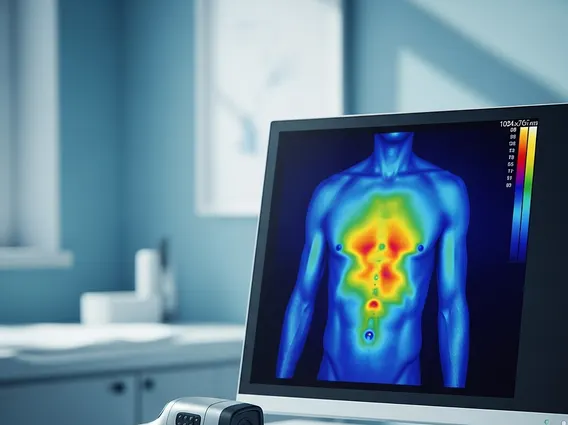
Infrared Thermography (IT) is a non-invasive, non-contact diagnostic imaging technique that measures and maps the skin surface temperature. It operates on the principle that metabolic activity and blood flow generate heat, which is then radiated from the body. This emitted infrared energy is detected by a specialized camera and converted into a visual image, known as a thermogram, where different colors represent varying temperatures. This method allows clinicians to observe temperature patterns that may indicate underlying physiological changes, making it a valuable tool in medical diagnostics.
Principles of Infrared Thermography
How infrared thermography works involves the detection of electromagnetic radiation in the infrared spectrum. All objects with a temperature above absolute zero emit infrared energy, and the human body is a significant emitter. An infrared camera contains sensors that capture this emitted radiation, which is then processed by a computer. The software translates the intensity of the infrared radiation into a visible spectrum, typically displayed as a color-coded image. Warmer areas often appear in red or white, while cooler areas may be shown in blue or purple. These thermal patterns reflect the physiological state of the tissues, as changes in blood flow, inflammation, or nerve activity can alter local skin temperature. The accuracy of the readings depends on factors like ambient temperature, patient preparation, and camera calibration.
Clinical Applications and Benefits
Infrared thermography applications span several medical disciplines, offering a unique perspective on physiological function. In oncology, it has been explored for its potential in detecting early signs of breast abnormalities by identifying areas of increased metabolic activity and angiogenesis associated with tumor growth. It is also used in pain management to localize sources of neuropathic or musculoskeletal pain by identifying areas of altered blood flow or inflammation. Other applications include assessing peripheral vascular disease, monitoring wound healing, and evaluating nerve damage.
The benefits of infrared thermography are numerous, particularly due to its non-invasive and radiation-free nature:
- Non-contact and Painless: The procedure involves no physical contact with the patient, making it comfortable and suitable for individuals of all ages, including those with sensitive conditions.
- Radiation-Free: Unlike X-rays or CT scans, IT does not use ionizing radiation, allowing for repeated examinations without cumulative exposure risks.
- Physiological Assessment: It provides functional information by detecting temperature changes that can precede anatomical changes, potentially aiding in earlier detection of certain conditions.
- Adjunctive Tool: It serves as a valuable adjunct to other diagnostic imaging modalities, offering complementary information that can enhance diagnostic accuracy.
This technology offers a safe and objective method for visualizing and quantifying thermal patterns, contributing to a more comprehensive understanding of a patient’s condition.


