Pyogenic Granuloma
Pyogenic Granuloma is a common, benign skin growth that appears as a small, reddish bump, often bleeding easily. While its name might suggest an infection, it is actually a vascular lesion characterized by an overgrowth of capillaries.
Key Takeaways
- Pyogenic Granuloma is a non-cancerous, rapidly growing vascular lesion.
- It often appears as a bright red, shiny, and easily bleeding bump on the skin or mucous membranes.
- Causes can include minor trauma, hormonal changes, or certain medications.
- Diagnosis is primarily clinical, often confirmed by biopsy to rule out other conditions.
- Treatment typically involves surgical removal, curettage, or laser therapy.
What is Pyogenic Granuloma?
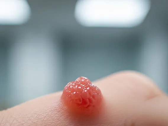
Pyogenic Granuloma is a benign vascular tumor that manifests as a rapidly growing, bright red to reddish-brown papule or nodule. Despite its misleading name, it is not infectious or granulomatous; rather, it is characterized by an excessive proliferation of capillaries and inflammatory cells. These lesions commonly appear on the skin, particularly on the head, neck, fingers, and trunk, but can also occur on mucous membranes such as the lips, gums, and nasal septum. It is a relatively common condition, affecting individuals of all ages, though it is frequently observed in children, young adults, and pregnant women, where hormonal fluctuations are thought to play a role.
The lesion typically presents as a soft, friable growth that bleeds easily with minimal trauma due to its rich vascularity. Its rapid growth can be alarming, often leading individuals to seek medical attention. While generally harmless, its appearance can sometimes mimic more serious conditions, necessitating accurate diagnosis.
Causes, Symptoms, and Diagnosis of Pyogenic Granuloma
The exact etiology of Pyogenic granuloma causes and symptoms is not fully understood, but several factors are believed to contribute to its development. Minor trauma, such as a cut or insect bite, is frequently implicated as a trigger, especially when the lesion appears at a site of injury. Hormonal changes, particularly during pregnancy, are also recognized as a significant factor, leading to the term “granuloma gravidarum” for lesions occurring in expectant mothers. Certain medications, including retinoids and some chemotherapy drugs, have also been associated with their emergence.
The primary symptoms are the appearance of a solitary, rapidly growing, reddish bump. Key characteristics include:
- Rapid growth, often appearing within weeks.
- Bright red, reddish-brown, or purplish color.
- Soft, friable texture, easily bleeding with minimal trauma.
- A moist or crusted surface, sometimes with a stalk-like base (pedunculated).
- Typically painless, unless traumatized or infected.
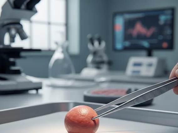
For Pyogenic granuloma diagnosis, a healthcare professional typically performs a clinical examination, observing the characteristic appearance and rapid growth. In many cases, a biopsy is recommended to confirm the diagnosis and differentiate it from other vascular lesions or more serious conditions like amelanotic melanoma or squamous cell carcinoma, which can have similar presentations. Histopathological examination reveals a lobular proliferation of capillaries within an edematous stroma.
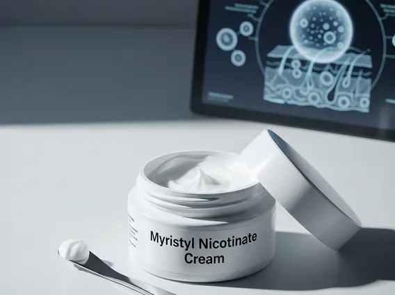
Pyogenic Granuloma Treatment Options
Effective Pyogenic granuloma treatment is crucial due to the lesion’s tendency to bleed and its potential for rapid growth. The most common and effective treatment is surgical excision, where the lesion is cut out and the base is cauterized to prevent recurrence. This method ensures complete removal and allows for histopathological examination of the tissue.
Another widely used approach is curettage and cauterization, involving scraping away the lesion with a curette followed by burning the base with an electric current to destroy any remaining vascular tissue. Other treatment modalities include laser therapy, particularly pulsed dye laser, which targets the blood vessels within the lesion and can be effective for smaller lesions or those in sensitive areas. Topical treatments, such as imiquimod cream or silver nitrate, may be used for very small lesions or as an adjunct, though they are generally less effective than surgical methods and carry a higher risk of recurrence. Recurrence rates vary depending on the treatment method and completeness of removal, but they can occur, especially if the lesion’s base is not adequately treated.