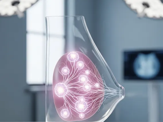
Pupil
The Pupil is a crucial component of the human eye, playing a vital role in vision. This small opening regulates the amount of light entering the eye, adapting constantly to various environmental conditions to optimize visual clarity.

Key Takeaways
- The Pupil is the opening in the center of the iris that controls the amount of light reaching the retina.
- Its size changes automatically through the pupillary reflex, governed by the autonomic nervous system.
- The function of the Pupil is to adapt vision to varying light levels, enhancing clarity in bright light and sensitivity in dim conditions.
- Pupil dilation can be influenced by factors such as light levels, emotional states, certain medications, and underlying medical conditions.
What is the Pupil?
The Pupil refers to the black circular opening located in the center of the iris of the eye. It is not a physical structure itself but rather an aperture that allows light to enter the eye and reach the retina. Its appearance as a black circle is due to the absorption of light by the tissues inside the eye, preventing it from being reflected back out.
The size of the Pupil is dynamic, constantly adjusting in response to light intensity and other stimuli. This involuntary adjustment is a critical part of the visual system, ensuring that the appropriate amount of light is processed for clear and comfortable vision.
Anatomy and Function of the Pupil
The anatomy of the human pupil is intrinsically linked to the iris, which is the colored part of the eye surrounding it. The iris contains two types of smooth muscles that control the pupil’s size: the sphincter pupillae and the dilator pupillae. The sphincter pupillae muscles are arranged in a circular pattern around the pupil and contract to make the pupil smaller (constriction). The dilator pupillae muscles are arranged radially, extending from the pupil to the edge of the iris, and they contract to make the pupil larger (dilation).
The function of the Pupil is to regulate the amount of light entering the eye. In bright light, the sphincter pupillae muscles contract, causing the pupil to constrict. This reduces the amount of light reaching the retina, preventing overstimulation and improving the depth of field, which enhances visual acuity. Conversely, in dim light, the dilator pupillae muscles contract, leading to pupil dilation. This allows more light to enter the eye, increasing the retina’s sensitivity and improving vision in low-light conditions.
The Pupil works through an involuntary reflex arc, primarily controlled by the autonomic nervous system. The parasympathetic nervous system mediates pupillary constriction, while the sympathetic nervous system mediates pupillary dilation. This intricate interplay ensures that the eye can rapidly adapt to changes in environmental illumination, optimizing visual performance across a wide range of light conditions.
What Causes Pupil Dilation?
Pupil dilation, medically known as mydriasis, is the widening of the pupil. Understanding what causes pupil dilation is important as it can be a normal physiological response or an indicator of various external influences or underlying medical conditions. This response is primarily mediated by the sympathetic nervous system, which triggers the dilator pupillae muscles to contract.
Several factors can lead to pupil dilation:
- Low Light Conditions: This is the most common physiological cause, as the pupils dilate to allow more light to reach the retina, improving vision in dim environments.
- Emotional Responses: Strong emotions such as fear, excitement, surprise, or even attraction can activate the sympathetic nervous system, leading to pupil dilation.
- Medications: Certain prescription and over-the-counter drugs can cause mydriasis. Examples include anticholinergics (like atropine), antihistamines, decongestants, and some antidepressant medications.
- Recreational Drugs: Stimulants (e.g., cocaine, amphetamines) and hallucinogens (e.g., LSD, psilocybin) are known to cause significant pupil dilation.
- Medical Conditions and Injuries: Conditions such as certain brain injuries, strokes, tumors, or optic nerve damage can affect the neurological pathways controlling pupil size, leading to dilation in one or both eyes. Adie’s pupil and Horner’s syndrome (in its acute phase) are also examples.
- Eye Drops for Examination: Ophthalmologists frequently use dilating eye drops during comprehensive eye exams to enlarge the pupil, allowing for a better view of the retina and optic nerve.
While temporary pupil dilation is often harmless, persistent or unexplained mydriasis, especially if accompanied by other symptoms like headache, vision changes, or eye pain, warrants medical evaluation to rule out serious underlying conditions.