Iobenguane Scan
An Iobenguane Scan is a specialized nuclear medicine imaging procedure used to detect and locate certain types of neuroendocrine tumors. This diagnostic tool plays a crucial role in the management of specific conditions by providing detailed insights into tumor presence and activity.
Key Takeaways
- An Iobenguane Scan uses a radioactive tracer (MIBG) to identify neuroendocrine tumors.
- It is primarily used for diagnosing and monitoring pheochromocytoma, paraganglioma, and neuroblastoma.
- The procedure involves an intravenous injection of the tracer followed by imaging sessions over one to three days.
- Results indicate areas of tracer uptake, helping physicians locate tumors and assess their metabolic activity.
- Interpretation of the scan requires expertise and is combined with other clinical information for accurate diagnosis.
What is an Iobenguane Scan?
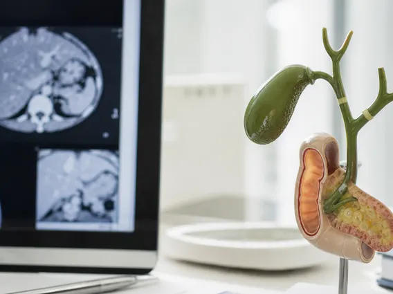
An Iobenguane Scan is a diagnostic imaging test that utilizes a small amount of a radioactive substance called meta-iodobenzylguanidine (MIBG) to visualize specific types of tumors. MIBG is structurally similar to norepinephrine, a hormone produced by nerve cells, and is selectively absorbed by certain neuroendocrine cells, particularly those found in pheochromocytomas, paragangliomas, and neuroblastomas. This allows the scan to pinpoint tumor locations that might not be visible with conventional imaging techniques.
The scan provides functional information, showing not just the anatomy but also the metabolic activity of the cells. This is particularly valuable for identifying tumors that produce catecholamines, which are hormones like adrenaline and noradrenaline. According to the National Cancer Institute, neuroendocrine tumors are a diverse group of tumors, and specialized imaging like the Iobenguane Scan helps in their precise characterization and staging.
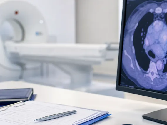
Iobenguane Scan Procedure and Uses
The Iobenguane scan procedure typically involves several steps over one to three days. Patients are often advised to stop certain medications, such as tricyclic antidepressants or decongestants, prior to the scan, as these can interfere with MIBG uptake. Thyroid blocking medication, such as potassium iodide, may also be administered to protect the thyroid gland from absorbing the radioactive iodine component of MIBG.
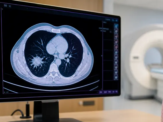
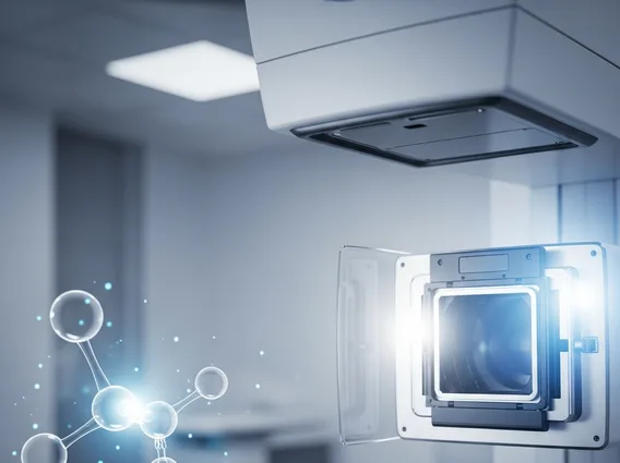
The procedure begins with the intravenous injection of the MIBG tracer. After the injection, there is a waiting period, usually a few hours to a day, to allow the tracer to circulate and accumulate in target tissues. Imaging sessions are then performed using a gamma camera, which detects the radiation emitted by the MIBG. Multiple imaging sessions may be conducted over subsequent days to capture optimal images as the tracer continues to distribute and clear from the body. These images often include single-photon emission computed tomography (SPECT) and computed tomography (CT) scans, which provide detailed 3D views.
The primary Iobenguane scan uses include:
- Diagnosis of Pheochromocytoma and Paraganglioma: These rare tumors originate from chromaffin cells and can produce excessive catecholamines. The Iobenguane Scan is highly effective in locating these tumors, whether they are in the adrenal glands (pheochromocytoma) or outside (paraganglioma).
- Staging and Monitoring of Neuroblastoma: A common childhood cancer, neuroblastoma often expresses the norepinephrine transporter, making it amenable to MIBG uptake. The scan helps determine the extent of the disease and monitor response to treatment.
- Identifying Metastases: It can detect if these tumors have spread to other parts of the body, which is crucial for treatment planning.
- Guiding Therapy: In some cases, a therapeutic form of MIBG (I-131 MIBG) can be used to treat these tumors, and the diagnostic scan helps identify suitable candidates for this targeted therapy.
Interpreting Iobenguane Scan Results
Understanding Iobenguane scan results explained is crucial for patient management. A nuclear medicine physician interprets the images, looking for areas of abnormal MIBG uptake. Normal uptake is typically seen in the salivary glands, liver, spleen, bladder, and heart. Any significant uptake outside these expected areas, or unusually intense uptake within them, can indicate the presence of a tumor.
A “positive” scan result means that areas of abnormal MIBG accumulation have been detected, suggesting the presence of a neuroendocrine tumor. The location, size, and intensity of uptake help determine the nature and extent of the disease. A “negative” scan result indicates no abnormal MIBG uptake, which typically suggests the absence of the targeted tumor type or that the tumor present does not absorb MIBG. It is important to note that a negative scan does not always rule out a tumor, as some tumors may not express the norepinephrine transporter.
The interpretation of Iobenguane Scan results is always considered in conjunction with a patient’s clinical symptoms, blood tests (e.g., catecholamine levels), and other imaging studies (e.g., MRI or CT scans). This comprehensive approach ensures the most accurate diagnosis and guides appropriate treatment strategies for conditions like pheochromocytoma, paraganglioma, and neuroblastoma.