Immunophenotyping
Immunophenotyping is a powerful laboratory technique used in medicine to identify and characterize cells based on the proteins expressed on their surface or inside them. This method provides critical insights into cell populations, particularly in the context of disease diagnosis and monitoring.
Key Takeaways
- Immunophenotyping identifies cells by detecting specific protein markers.
- It primarily uses techniques like flow cytometry with fluorescent antibodies.
- This method is crucial for diagnosing and classifying blood cancers such as leukemias and lymphomas.
- It aids in monitoring disease progression and assessing treatment effectiveness.
- The insights gained from immunophenotyping guide targeted therapies and improve patient outcomes.
What is Immunophenotyping?
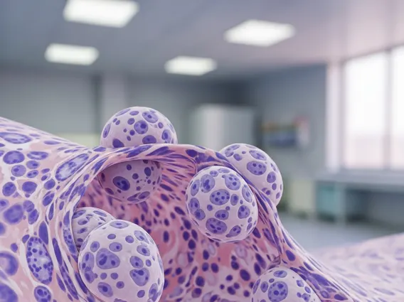
Immunophenotyping refers to a laboratory technique used to identify and characterize cells based on the expression of specific proteins, known as antigens or markers, on their surface or within their cytoplasm. These markers act like unique cellular fingerprints, allowing scientists and clinicians to distinguish different cell types and their developmental stages. The immunophenotyping meaning is rooted in its ability to reveal the precise identity and functional state of cells, which is particularly vital in understanding complex biological processes and disease states. This technique is fundamental in various medical fields, especially in hematology, immunology, and oncology, where accurate cell identification is paramount for diagnosis and treatment. Essentially, immunophenotyping explained involves using antibodies that bind specifically to these cellular markers, making them detectable.
How Does Immunophenotyping Work?
The process of immunophenotyping primarily relies on the use of antibodies, which are proteins that specifically recognize and bind to particular antigens on cells. These antibodies are typically conjugated with fluorescent dyes, allowing the tagged cells to be detected and analyzed. The most common technology employed for this purpose is flow cytometry.
Here’s a simplified overview of how immunophenotyping works using flow cytometry:
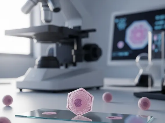
- Sample Preparation: A biological sample, such as blood, bone marrow, or tissue biopsy, is collected and processed to obtain a suspension of individual cells.
- Antibody Staining: The prepared cells are incubated with a panel of fluorescently labeled antibodies. Each antibody is designed to bind to a specific cellular marker.
- Flow Cytometry Analysis: The stained cells are then passed through a flow cytometer in a single-file stream. As each cell passes through a laser beam, the fluorescent dyes are excited, emitting light at specific wavelengths.
- Data Acquisition and Interpretation: Detectors measure the scattered light and emitted fluorescence from each cell. This data is then analyzed by specialized software to create plots that show the expression patterns of different markers, allowing for the identification and quantification of various cell populations.
This precise method enables the differentiation of normal cells from abnormal or malignant cells, providing a detailed cellular profile.
Clinical Applications and Importance of Immunophenotyping
Immunophenotyping plays a critical role in modern clinical diagnostics, offering invaluable insights that guide patient management, particularly in hematologic malignancies and immune disorders. Its primary clinical applications include:
- Diagnosis and Classification of Leukemias and Lymphomas: Immunophenotyping is indispensable for accurately diagnosing and subtyping various types of leukemia and lymphoma. By identifying specific marker combinations, clinicians can distinguish between acute myeloid leukemia (AML), acute lymphoblastic leukemia (ALL), and different types of non-Hodgkin lymphoma, which often look similar under a microscope but require distinct treatment approaches.
- Prognostic Assessment: The presence or absence of certain markers can provide crucial prognostic information, indicating how aggressive a disease might be and its likely response to therapy. This helps in risk stratification and tailoring treatment intensity.
- Monitoring Minimal Residual Disease (MRD): After treatment, immunophenotyping can detect very small numbers of remaining cancer cells (MRD) that are undetectable by conventional methods. MRD monitoring is vital for assessing treatment effectiveness and predicting relapse, allowing for timely intervention.
- Diagnosis of Primary Immunodeficiency Disorders: It helps identify deficiencies or abnormalities in specific immune cell populations, aiding in the diagnosis of various primary immunodeficiency disorders.
The importance of immunophenotyping cannot be overstated. According to the World Health Organization (WHO), accurate and timely diagnosis is a cornerstone of effective cancer control, and advanced diagnostic tools like immunophenotyping significantly contribute to this goal by enabling precise disease classification. This precision allows for the selection of targeted therapies, leading to improved patient outcomes and reduced treatment-related toxicities.