
Radiofrequency Ablation
Radiofrequency Ablation (RFA) is a minimally invasive medical procedure that uses heat generated by radio waves to destroy specific tissues, most commonly tumors or nerve pathways causing chronic pain. It offers a targeted approach for various medical conditions, providing an alternative to more invasive surgical interventions.
Key Takeaways
- Radiofrequency Ablation (RFA) is a minimally invasive procedure using heat from radio waves to destroy targeted tissues.
- It is commonly used to treat cancerous tumors in organs like the liver, kidney, and lung, as well as to alleviate chronic pain.
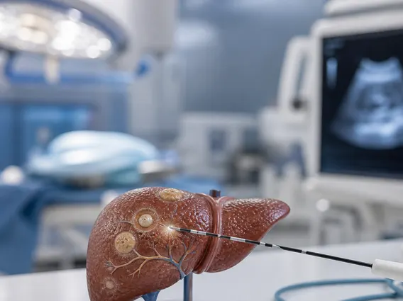
- The procedure involves inserting a thin needle electrode into the target area, guided by imaging, to deliver radiofrequency energy.
- Benefits include reduced recovery time, minimal scarring, and effectiveness for localized conditions.
- Potential risks, though generally low, include pain, bleeding, infection, and damage to surrounding healthy tissue.
What is Radiofrequency Ablation?
Radiofrequency Ablation (RFA) refers to a medical procedure that employs high-frequency electrical currents to generate heat, which in turn destroys abnormal tissue. This technique is primarily used in oncology to treat tumors in organs such as the liver, kidney, lung, and bone. Beyond cancer treatment, RFA is also a recognized method for managing chronic pain, particularly in conditions affecting the spine or joints, by ablating nerve pathways responsible for transmitting pain signals. The principle behind RFA is the precise delivery of thermal energy to a localized area, causing cellular death within the targeted tissue while minimizing damage to surrounding healthy structures. This minimally invasive approach has become a valuable option for patients who may not be candidates for traditional surgery or as an adjunct therapy.
How Radiofrequency Ablation Works and Procedure Details
How radiofrequency ablation works involves the insertion of a specialized needle electrode directly into the targeted tissue. This electrode is connected to a radiofrequency generator, which emits alternating electrical currents. As these currents pass through the tissue, they cause ions within the cells to vibrate rapidly, generating frictional heat. When the tissue temperature reaches approximately 60-100°C, the cells undergo coagulative necrosis, effectively destroying the abnormal tissue. The precise placement of the electrode is crucial and is typically achieved using imaging guidance such as ultrasound, CT scans, or MRI, allowing the physician to visualize the target area in real-time.
The radiofrequency ablation procedure details generally follow a structured approach:
- Preparation: The patient is typically given local anesthesia, conscious sedation, or sometimes general anesthesia, depending on the location and complexity of the procedure. Imaging guidance is established to map the target area.
- Insertion: A thin, hollow needle electrode is carefully advanced through the skin and into the target lesion or nerve, guided by the chosen imaging modality.
- Energy Delivery: Once the electrode is correctly positioned, radiofrequency energy is delivered through its tip. The duration and intensity of the energy delivery are precisely controlled to achieve adequate tissue destruction.
- Monitoring: Throughout the procedure, the physician monitors the patient’s vital signs and the ablation zone using imaging to ensure safety and effectiveness.
- Post-Procedure: After the ablation, the electrode is withdrawn, and a small bandage is applied. Patients are typically observed for a few hours before discharge, with specific post-procedure instructions provided.
Benefits and Risks of Radiofrequency Ablation
Radiofrequency ablation benefits include its minimally invasive nature, which often translates to shorter hospital stays, quicker recovery times, and less post-procedural pain compared to open surgery. It can be performed on an outpatient basis for many conditions, allowing patients to return to their daily activities sooner. RFA is also a viable option for patients who are not suitable for surgery due to age, comorbidities, or the location of the lesion. Furthermore, it offers a targeted treatment, preserving surrounding healthy tissue. For instance, in liver cancer, RFA has shown comparable efficacy to surgical resection for small tumors, with lower morbidity rates according to studies cited by organizations like the National Cancer Institute.
However, like any medical intervention, there are radiofrequency ablation risks that patients should be aware of. While generally safe, potential complications can include:
- Pain: Discomfort at the ablation site, which is usually manageable with medication.
- Bleeding or Hematoma: Bruising or blood collection at the needle insertion site.
- Infection: Though rare, there is a risk of infection at the site or within the treated area.
- Damage to Adjacent Structures: In some cases, nearby healthy tissues, nerves, or organs may be inadvertently affected by the heat.
- Pneumothorax: A collapsed lung, particularly when treating lung lesions.
- Skin Burns: Rare, but possible at the electrode insertion site.
Patients should discuss these potential benefits and risks thoroughly with their healthcare provider to determine if RFA is the most appropriate treatment option for their specific condition.



















