Ct Scan
A Computed Tomography (CT) scan is a sophisticated medical imaging technique that utilizes X-rays and computer processing to create detailed cross-sectional images of the body. This non-invasive diagnostic tool plays a crucial role in modern medicine, aiding in the detection and diagnosis of a wide range of conditions.
Key Takeaways
- A CT scan uses specialized X-ray equipment and computer technology to produce detailed, cross-sectional images of internal organs, bones, soft tissue, and blood vessels.
- It is a vital diagnostic tool for identifying various medical conditions, including injuries, diseases, and abnormalities, by providing more detail than conventional X-rays.
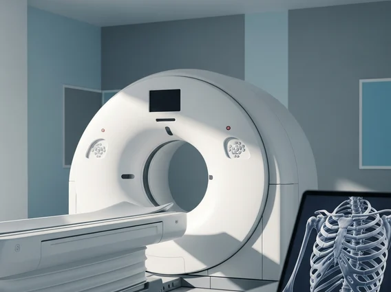
- The procedure is generally quick, painless, and involves the patient lying on a table that moves through a donut-shaped scanner.
- While CT scans involve exposure to ionizing radiation, the benefits for accurate diagnosis often outweigh the potential risks, which are carefully managed by medical professionals.
- Contrast agents may be used to enhance image clarity, though they carry a small risk of allergic reactions or kidney issues in some individuals.
What is a CT Scan (Computed Tomography)?
A Computed Tomography (CT) scan, often simply referred to as a CT scan, is a powerful diagnostic imaging procedure that combines a series of X-ray images taken from different angles around your body. A computer then processes these images to create detailed cross-sectional slices, or “tomographic” images, of the bones, blood vessels, and soft tissues inside your body. Unlike a conventional X-ray, which produces a single 2D image, a CT scan provides much more comprehensive and detailed views, allowing medical professionals to see internal structures with remarkable clarity. This advanced imaging capability makes it an indispensable tool for diagnosing and monitoring numerous medical conditions.
How CT Scans Work and Their Medical Applications
The process of a CT scan involves a patient lying on a motorized table that slides into a large, donut-shaped machine called a gantry. Inside the gantry, an X-ray tube rotates around the patient, emitting a narrow beam of X-rays. On the opposite side of the patient, specialized detectors measure the X-rays that pass through the body. As the X-ray tube and detectors rotate, they capture thousands of individual images from various angles. A powerful computer then takes this vast amount of data and reconstructs it into detailed cross-sectional images, which can be viewed in 2D or even 3D. Sometimes, a contrast material, administered orally or intravenously, is used to highlight specific areas like blood vessels, organs, or tumors, making them more visible on the images.
The uses of CT scan are extensive across various medical specialties due to its ability to provide intricate details of internal structures. Common applications include:
- Diagnosing Injuries: Identifying fractures, internal bleeding, and organ damage after trauma.
- Detecting Diseases: Locating tumors, infections, blood clots, and other abnormalities in organs like the lungs, liver, kidneys, and brain.
- Guiding Procedures: Assisting doctors during biopsies, radiation therapy, and surgical planning by providing precise anatomical guidance.
- Monitoring Conditions: Tracking the progression of diseases like cancer or heart disease, and evaluating the effectiveness of treatments.
- Assessing Vascular Issues: Visualizing blood vessels to detect aneurysms, blockages, or other vascular conditions.
Potential Risks and Safety Considerations
While CT scans are invaluable diagnostic tools, it is important to be aware of the potential risks of CT scan procedures. The primary concern is exposure to ionizing radiation. Although the amount of radiation from a single CT scan is generally low, cumulative exposure from multiple scans over time can slightly increase the lifetime risk of cancer. Medical professionals adhere to the “As Low As Reasonably Achievable” (ALARA) principle, ensuring that scans are only performed when medically necessary and using the lowest possible radiation dose to obtain diagnostic quality images. For pregnant women, CT scans are generally avoided unless absolutely critical, as radiation exposure can pose risks to the developing fetus.
Another consideration involves the use of contrast agents. While generally safe, some individuals may experience allergic reactions, ranging from mild itching or hives to more severe anaphylaxis. Patients with pre-existing kidney conditions may also be at a higher risk of kidney damage from certain intravenous contrast dyes. It is crucial for patients to inform their healthcare provider about any allergies, kidney problems, or other medical conditions before undergoing a CT scan with contrast. Overall, the diagnostic benefits of a CT scan in providing critical information for patient care typically outweigh these potential risks when the procedure is medically indicated and performed under appropriate supervision.