Isointense
In the field of medical imaging, particularly magnetic resonance imaging (MRI), the term isointense is crucial for describing and interpreting tissue characteristics. It refers to a signal intensity that is similar to, or indistinguishable from, the surrounding tissues or a reference tissue.
Key Takeaways
- Isointense describes a signal intensity in medical imaging that matches that of surrounding tissues.
- In MRI, isointense signal characteristics mean a lesion or structure appears similar in brightness to its environment.
- This similarity can make detection challenging, often requiring comparison across different MRI sequences.
- Understanding isointense vs hypointense signals is fundamental for accurate diagnosis, as hypointense signals appear darker.
What is Isointense?
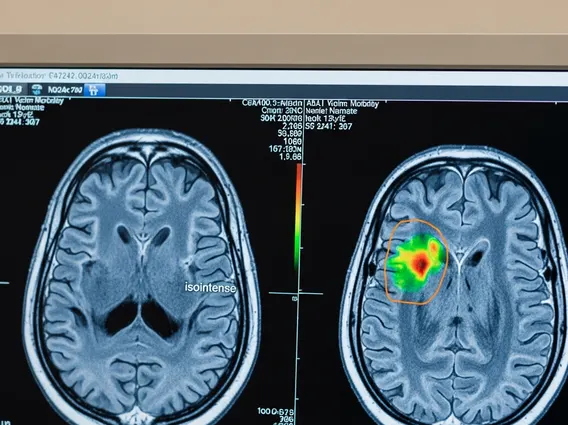
Isointense refers to a phenomenon observed in medical imaging, primarily magnetic resonance imaging (MRI), where a specific tissue or lesion exhibits a signal intensity that is comparable to, or the same as, the signal intensity of the adjacent or surrounding normal tissues. This means that on an MRI scan, the area in question appears neither significantly brighter (hyperintense) nor significantly darker (hypointense) than its immediate environment. The concept of isointensity is vital for radiologists and clinicians as it helps in characterizing abnormalities and differentiating them from healthy structures.
When a structure is described as isointense, it implies that its physical properties, such as water content, fat content, or cellular density, are similar to those of the surrounding tissues under the specific imaging sequence used. This can sometimes make it challenging to identify lesions, as they may blend in with the normal anatomy. Therefore, multiple imaging sequences and careful comparison are often necessary to detect and characterize isointense findings.
Isointense Signal Characteristics in MRI
The isointense signal characteristics in MRI are determined by how different tissues interact with the magnetic fields and radiofrequency pulses during the scan. The isointense mri definition hinges on the fact that the relaxation times (T1 and T2) of the tissue of interest are similar to those of the reference tissue. For instance, on a T1-weighted image, a lesion might appear isointense if its T1 relaxation time is similar to that of the surrounding brain parenchyma. Similarly, on a T2-weighted image, isointensity suggests comparable T2 relaxation times.
Several factors can contribute to a tissue appearing isointense. These include:
- Water Content: Tissues with similar water content to their surroundings often appear isointense.
- Cellular Density: Areas with comparable cellularity to adjacent normal tissue can also exhibit isointensity.
- Fat Content: The presence and distribution of fat can influence signal intensity, leading to isointense appearances if matched with surrounding fatty tissues.
- Pathological Changes: Early-stage lesions or certain types of tumors may initially appear isointense, making them difficult to detect without contrast agents or advanced imaging techniques.
Radiologists often analyze signal intensity across various MRI sequences (e.g., T1-weighted, T2-weighted, FLAIR, diffusion-weighted imaging) to fully characterize a lesion. A finding that is isointense on one sequence might become hyperintense or hypointense on another, providing crucial diagnostic information.
Isointense vs. Hypointense Signals
Understanding the distinction between isointense vs hypointense signals is fundamental for accurate interpretation of medical images. While isointense signals blend with their surroundings, hypointense signals stand out by appearing darker. Hypointense refers to a signal intensity that is lower than that of the surrounding tissues or a reference tissue on an imaging scan. This typically means the area appears darker on the image.
The differences in signal intensity are critical for identifying and characterizing various pathologies. For example, on T1-weighted MRI, cerebrospinal fluid (CSF) typically appears hypointense (dark), while on T2-weighted images, it is hyperintense (bright). A lesion that is isointense on one sequence but hypointense on another provides specific clues about its composition and nature.
| Characteristic | Isointense Signal | Hypointense Signal |
|---|---|---|
| Appearance on Image | Similar brightness to surrounding tissue | Darker than surrounding tissue |
| Signal Intensity | Comparable to adjacent structures | Lower than adjacent structures |
| Interpretation | May indicate similar tissue composition; can be challenging to detect lesions | Often indicates lower water content, calcification, fibrosis, or certain fluid collections |
| Diagnostic Implication | Requires multi-sequence comparison; may need contrast for lesion detection | Easily distinguishable; often indicative of specific pathological processes |
In summary, the ability to differentiate between isointense and hypointense signals is a cornerstone of diagnostic imaging. While isointensity can sometimes obscure subtle abnormalities, hypointensity often highlights areas of significant change, guiding clinicians toward an accurate diagnosis and appropriate patient management.