Immunohistochemistry
Immunohistochemistry is a powerful laboratory technique that plays a pivotal role in medical diagnostics and research. It allows scientists and clinicians to visualize the precise location of specific proteins (antigens) within tissue samples, providing critical insights into various diseases.

Key Takeaways
- Immunohistochemistry (IHC) is a diagnostic technique using antibodies to detect specific antigens in tissue.
- It relies on the principle of highly specific antibody-antigen binding, visualized through enzymatic reactions or fluorescence.
- The immunohistochemistry staining procedure involves meticulous steps from tissue preparation to microscopic examination.
- IHC is indispensable in pathology for accurate disease diagnosis, particularly in classifying tumors and guiding treatment strategies.
- It helps identify prognostic markers and predict patient response to targeted therapies, advancing personalized medicine.
What is Immunohistochemistry (IHC)?
Immunohistochemistry (IHC) is a sophisticated laboratory technique that combines principles from immunology and histology to detect specific antigens (proteins) in cells and tissues. This method utilizes the highly specific binding of antibodies to their corresponding antigens, allowing for the visualization of these molecular targets within their cellular context. By identifying the presence, absence, or overexpression of particular proteins, IHC provides crucial insights into cellular processes, disease progression, and therapeutic targets. It has become an indispensable tool in diagnostic pathology, particularly in oncology, where precise identification of cellular markers is critical for accurate diagnosis and patient management.
Immunohistochemistry Principles and Staining Procedure
The core of immunohistochemistry principle applications lies in the specific interaction between an antibody and its target antigen. When a tissue sample containing the antigen is exposed to a primary antibody designed to bind specifically to that antigen, a complex forms. To make this binding visible, a secondary antibody, often conjugated with an enzyme (like horseradish peroxidase or alkaline phosphatase) or a fluorescent dye, is introduced. This secondary antibody binds to the primary antibody, and when a suitable substrate is added, the enzyme catalyzes a reaction that produces a colored precipitate or a fluorescent signal at the site of the antigen, which can then be observed under a microscope.
The immunohistochemistry staining procedure is a multi-step process designed to ensure optimal antigen detection and minimal background noise. Key steps typically include:
- Tissue Preparation: Formalin-fixed, paraffin-embedded (FFPE) tissue sections are commonly used, requiring deparaffinization and rehydration.
- Antigen Retrieval: Often necessary for FFPE tissues to unmask antigens altered by fixation, making them accessible to antibodies.
- Blocking: Non-specific binding sites are blocked to prevent antibodies from binding to unintended targets, reducing background staining.
- Primary Antibody Incubation: The tissue section is incubated with the primary antibody specific to the target antigen.
- Washing: Excess primary antibody is removed.
- Secondary Antibody Incubation: A labeled secondary antibody, which binds to the primary antibody, is applied.
- Washing: Excess secondary antibody is removed.
- Detection: A chromogen (for enzymatic labels) or fluorophore is applied to visualize the antigen-antibody complex.
- Counterstaining: A contrasting stain (e.g., hematoxylin) is used to highlight cellular structures, providing morphological context.
- Mounting and Visualization: The stained slide is mounted and examined under a light or fluorescent microscope.
This meticulous process ensures accurate and reliable detection of cellular antigens, providing valuable diagnostic information.
Applications of Immunohistochemistry in Diagnostics
Immunohistochemistry has revolutionized diagnostic pathology, offering unparalleled precision in identifying and characterizing various diseases. Its most significant impact is in oncology, where it is routinely used for:
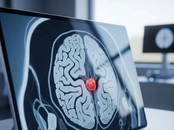
- Tumor Diagnosis and Classification: IHC helps pathologists differentiate between various types of cancers, such as distinguishing carcinomas from lymphomas or sarcomas, and further subclassify them based on specific protein expression patterns. For instance, different types of breast cancer can be identified by their expression of estrogen receptor (ER), progesterone receptor (PR), and HER2.
- Determining Primary Site of Metastasis: When a metastatic tumor is found, IHC can help identify the likely origin of the primary tumor, which is crucial for guiding appropriate treatment strategies.
- Prognostic and Predictive Markers: IHC identifies markers that predict disease aggressiveness (prognostic markers) or the likelihood of response to specific therapies (predictive markers). For example, PD-L1 expression is assessed via IHC to determine eligibility for certain immunotherapies in various cancers.
Beyond oncology, IHC is also valuable in diagnosing infectious diseases by detecting microbial antigens, identifying neurological disorders through specific protein aggregates, and characterizing renal diseases. The ability of IHC to provide both molecular and morphological information within the tissue context makes it an indispensable tool for accurate diagnosis and personalized medicine.