H And E Staining
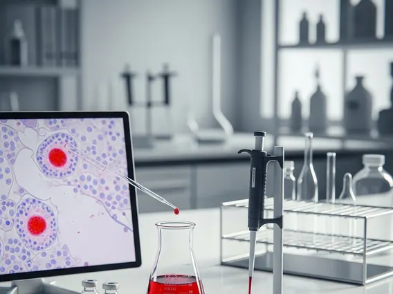
H&E staining, or Hematoxylin and Eosin staining, is a fundamental technique in histology and pathology, widely used for examining tissue morphology. It provides critical insights into cellular structures, aiding in the diagnosis of various diseases.
Key Takeaways
- H&E staining is a cornerstone diagnostic tool in pathology, revealing tissue architecture and cellular details.
- The technique uses two dyes: hematoxylin, which stains acidic components blue/purple, and eosin, which stains basic components pink/red.
- It helps pathologists identify normal and abnormal cellular structures, crucial for diagnosing conditions like cancer, inflammation, and infections.
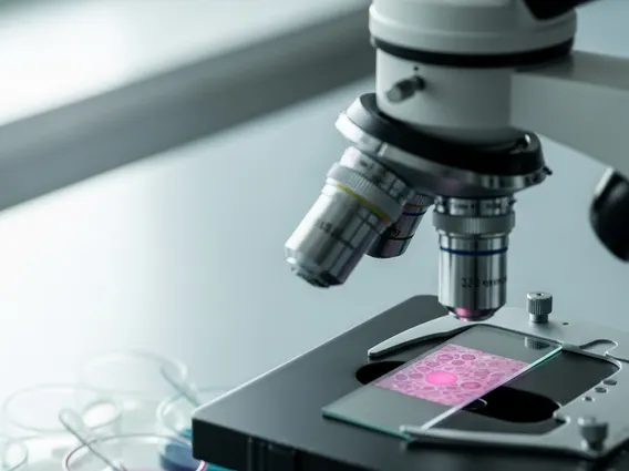
- The procedure involves tissue preparation, sectioning, staining, and mounting, resulting in slides that are examined under a microscope.
- Interpreting H&E stained slides requires expertise to differentiate various cell types and tissue components based on their color and morphology.
What is H&E (Hematoxylin and Eosin) Staining?
H&E staining, short for Hematoxylin and Eosin staining, is a widely used histological staining method that highlights different tissue components with contrasting colors. This technique is indispensable in medical diagnostics, particularly in surgical pathology, as it allows pathologists to visualize the microscopic structure of tissues and cells. The H&E staining explanation lies in its ability to differentiate between acidic and basic cellular components, providing a clear morphological overview.
The method employs two primary dyes: hematoxylin and eosin. Hematoxylin is a basic dye that stains basophilic structures (acidic components) blue or purple, primarily targeting the cell nuclei, ribosomes, and rough endoplasmic reticulum. Eosin, an acidic dye, stains acidophilic structures (basic components) pink or red, highlighting the cytoplasm, collagen, muscle fibers, and red blood cells. This differential staining creates a clear contrast, making it easier to identify various cell types and tissue organization.
How H&E Staining Works: Principles and Procedure
The process of how H&E staining works involves a series of precise steps to prepare tissue samples for microscopic examination. The underlying principles rely on the chemical properties of the dyes and their affinity for specific cellular components. Hematoxylin, often used with a mordant like aluminum salts, forms a complex that binds to negatively charged nucleic acids in the nucleus, giving it a characteristic blue-purple color. Eosin, being negatively charged, binds to positively charged proteins in the cytoplasm and extracellular matrix, resulting in various shades of pink.
The typical procedure for H&E staining involves several key stages:
- Tissue Fixation: Preserving tissue structure, usually with formalin.
- Processing and Embedding: Dehydrating the tissue and embedding it in paraffin wax to provide support for sectioning.
- Sectioning: Cutting the paraffin block into very thin slices (typically 3-5 micrometers) using a microtome.
- Deparaffinization and Rehydration: Removing the wax and rehydrating the tissue sections for aqueous staining.
- Hematoxylin Staining: Immersing the slides in hematoxylin solution to stain the nuclei.
- Eosin Staining: Immersing the slides in eosin solution to stain the cytoplasm and extracellular matrix.
- Dehydration and Clearing: Removing water and making the tissue transparent using alcohol and xylene.
- Mounting: Placing a coverslip over the stained tissue with a mounting medium for permanent preservation.
These steps ensure optimal visualization of cellular and tissue morphology, making H&E staining uses and principles foundational to diagnostic pathology.
Interpreting H&E Stained Slides: Uses and Significance
Interpreting H&E stained slides is a critical skill for pathologists, allowing them to diagnose diseases by observing cellular and tissue changes. The distinct coloration provided by hematoxylin and eosin helps differentiate various components: nuclei appear blue/purple, cytoplasm ranges from pale pink to intense red, and extracellular matrix components like collagen stain pink. This visual contrast is essential for identifying normal tissue architecture versus pathological alterations.
The primary use of H&E staining is in the diagnosis of a wide array of conditions, including:
| Component | H&E Staining Color | Significance |
|---|---|---|
| Nuclei | Blue/Purple | Indicates genetic material; changes in size, shape, or chromatin pattern can suggest malignancy or infection. |
| Cytoplasm | Pink/Red | Reflects protein content and organelles; variations can indicate cellular activity, damage, or differentiation. |
| Collagen/Connective Tissue | Pink | Provides structural support; alterations can be seen in fibrosis, inflammation, or tumors. |
| Red Blood Cells | Bright Red | Presence and location can indicate hemorrhage or vascular changes. |
By understanding H&E stained slides, pathologists can detect subtle changes in cell morphology, nuclear-to-cytoplasmic ratio, presence of abnormal cells, and tissue organization. This enables them to classify tumors, identify inflammatory processes, detect infectious agents, and assess tissue viability, making H&E staining an indispensable tool in clinical diagnostics and medical research.