Cm Avm Syndrome
Cm Avm Syndrome refers to a specific type of cerebral arteriovenous malformation, a rare condition characterized by an abnormal tangle of blood vessels in the brain. This article provides a comprehensive overview of this syndrome, covering its nature, symptoms, causes, diagnosis, and management.

Key Takeaways
- Cm Avm Syndrome is a rare, congenital vascular anomaly in the brain where arteries connect directly to veins without capillaries.
- Symptoms often include headaches, seizures, and neurological deficits, varying based on the malformation’s size and location.
- The exact cause is unknown, but it is believed to be present from birth.
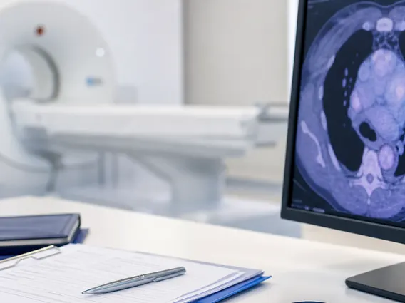
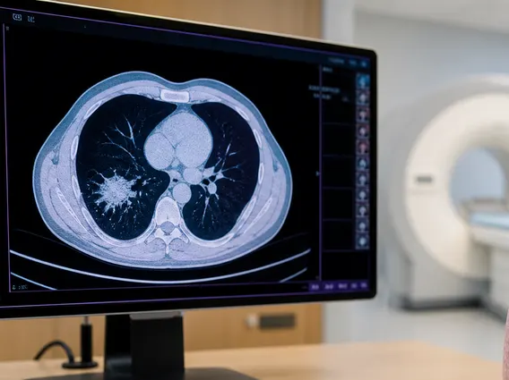
- Diagnosis relies on advanced imaging techniques such as MRI, CT scans, and cerebral angiography.
- Management options range from observation to surgical removal, embolization, or radiosurgery, tailored to the individual case.
What is Cm Avm Syndrome?
Cm Avm Syndrome is a medical condition characterized by a specific type of Cerebral Arteriovenous Malformation (AVM). An AVM is an abnormal tangle of blood vessels where arteries connect directly to veins, bypassing the normal capillary network. This direct connection creates a high-pressure shunt, as arteries carry high-pressure blood directly into lower-pressure veins, which can weaken the vessel walls over time. While AVMs can occur anywhere in the body, those in the brain, known as cerebral AVMs, are particularly concerning due to their potential to cause serious neurological complications.
The prevalence of cerebral AVMs is relatively low, affecting less than 1% of the general population. According to the American Stroke Association, approximately 300,000 Americans have an AVM, with about 1% of the general population being affected by some form of AVM. Cm Avm Syndrome, as a specific manifestation, requires careful medical attention due to the risks associated with hemorrhage and neurological impairment.
Symptoms and Causes of Cerebral Arteriovenous Malformation (AVM)
The symptoms of a Cerebral AVM can vary widely depending on the size, location, and whether it has bled. Many individuals with an AVM may not experience any symptoms until the malformation ruptures, leading to a brain hemorrhage. However, some may present with chronic symptoms. Common symptoms associated with cerebral AVMs include:
- Severe headaches, often localized to one side of the head.
- Seizures, which can range from focal to generalized.
- Neurological deficits such as muscle weakness, numbness, or paralysis on one side of the body.
- Problems with coordination, balance, or speech.
- Vision disturbances or double vision.
- Confusion or difficulty with memory.
The exact causes of cerebral AVMs are not fully understood, but they are generally considered to be congenital, meaning they are present at birth. They are not typically inherited, and most cases occur sporadically without a clear genetic link. Researchers believe that AVMs develop during fetal development when the blood vessels are forming. Factors that might contribute to their formation are still under investigation, but they are not usually linked to environmental exposures or lifestyle choices.
Diagnosis and Management of Cm Avm Syndrome
The Arteriovenous Malformation diagnosis process typically begins when a patient presents with symptoms suggestive of a neurological issue or when an AVM is incidentally discovered during imaging for another condition. Advanced imaging techniques are crucial for confirming the presence of an AVM, determining its exact location, size, and characteristics, and assessing any associated bleeding or brain damage. Key diagnostic tools include:
| Diagnostic Method | Description |
|---|---|
| Magnetic Resonance Imaging (MRI) | Provides detailed images of brain structures and can detect AVMs and any associated hemorrhage or edema. |
| Computed Tomography (CT) Scan | Useful for quickly identifying acute bleeding in the brain, often the first scan performed in emergency situations. |
| Cerebral Angiography | Considered the gold standard for AVM diagnosis, involving injecting contrast dye into blood vessels to visualize the AVM’s precise architecture and blood flow patterns. |
Management of Cm Avm Syndrome is highly individualized and depends on several factors, including the patient’s age, overall health, the AVM’s size and location, and the presence of symptoms or prior hemorrhage. Treatment options aim to prevent rupture and alleviate symptoms. These may include:
- Observation: For asymptomatic AVMs with low risk of rupture, a watchful waiting approach may be adopted.
- Surgical Resection: Direct surgical removal of the AVM, often the preferred method for accessible malformations.
- Endovascular Embolization: A minimally invasive procedure where a catheter is used to deliver materials that block blood flow to the AVM.
- Stereotactic Radiosurgery: Uses highly focused radiation beams to damage the AVM vessels, causing them to gradually close off over time.
A multidisciplinary team, including neurosurgeons, neurologists, and interventional neuroradiologists, typically collaborates to determine the most appropriate treatment strategy for each patient.