Fluorescence Microscopy
Fluorescence Microscopy is a powerful optical imaging technique that utilizes the phenomenon of fluorescence to visualize specimens, offering high sensitivity and specificity in biological and medical research. It allows scientists to observe specific molecules, structures, and processes within cells and tissues with remarkable detail.

Key Takeaways
- Fluorescence Microscopy uses fluorescent dyes or proteins to illuminate specific structures, enabling detailed visualization of biological samples.
- The technique relies on the principle of fluorophores absorbing light at one wavelength and emitting it at a longer, different wavelength.
- Key components include an excitation light source, filters, a dichroic mirror, and a detector to capture emitted light.
- It is widely applied in medical research for cell biology, pathology, diagnostics, and understanding disease mechanisms.
- This method provides high contrast and specificity, making it invaluable for studying complex biological systems.
What is Fluorescence Microscopy?
Fluorescence Microscopy is an advanced light microscopy technique that leverages the intrinsic or extrinsic fluorescent properties of a sample to generate an image. Unlike traditional light microscopy, which relies on light absorption or scattering, fluorescence microscopy uses fluorophores—molecules that absorb light at a specific wavelength (excitation) and then emit light at a longer, different wavelength (emission). This process allows for the visualization of specific components within a complex biological sample, such as cells or tissues, with high contrast and resolution.
This method is particularly valuable in medical and biological fields because it enables researchers to tag and observe particular proteins, organelles, or genetic material without interfering with the surrounding structures. By selecting appropriate fluorophores, scientists can label multiple targets simultaneously, providing a comprehensive view of cellular architecture and molecular interactions.
How Fluorescence Microscopy Works: Principles and Techniques
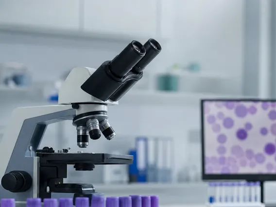
The fundamental fluorescence microscopy principles involve the interaction of light with fluorescent molecules. A typical fluorescence microscope consists of several key components that work in concert to achieve imaging. An intense light source, such as a mercury lamp, xenon lamp, or laser, emits light that passes through an excitation filter. This filter selects only the specific wavelength of light required to excite the fluorophores in the sample.
The filtered excitation light then reflects off a dichroic mirror and is focused onto the specimen by an objective lens. Upon absorbing the excitation light, the fluorophores in the sample become excited and subsequently emit light at a longer wavelength. This emitted fluorescent light passes back through the objective lens, travels through the dichroic mirror (which allows the longer wavelength light to pass), and then through an emission filter. The emission filter blocks any remaining excitation light, ensuring that only the fluorescent signal reaches the detector, typically a camera or the observer’s eye, to form a high-contrast image.
The process can be summarized by these steps:
- Excitation: A specific wavelength of light illuminates the fluorophore-labeled sample.
- Absorption: Fluorophores absorb the excitation light, moving to a higher energy state.
- Emission: Fluorophores return to their ground state, emitting light at a longer wavelength.
- Detection: Emitted light is captured and processed to form an image, while excitation light is blocked.
Applications of Fluorescence Microscopy in Medical Research
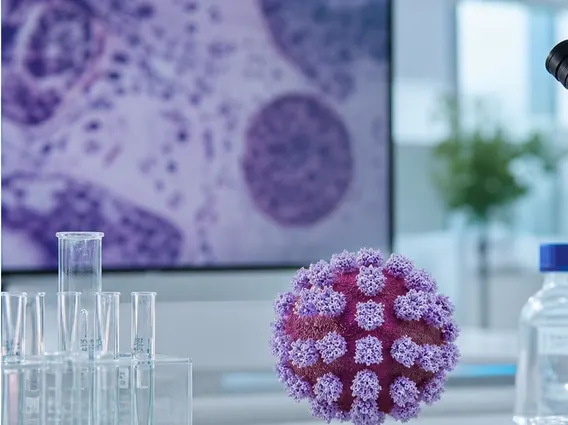
The applications of fluorescence microscopy are extensive and transformative across various domains of medical research, diagnostics, and drug discovery. Its ability to visualize specific cellular components with high sensitivity makes it an indispensable tool for understanding disease mechanisms and developing new therapies. For instance, in oncology, fluorescence microscopy is used to study tumor cell growth, metastasis, and the efficacy of anti-cancer drugs by labeling specific proteins involved in these processes.
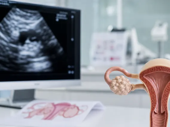
In cell biology, researchers utilize this technique to observe dynamic cellular processes such as protein trafficking, cell division, and immune responses. It is also crucial in pathology for diagnosing infectious diseases and identifying specific biomarkers in tissue samples, aiding in the classification and prognosis of various conditions. The development of advanced techniques like super-resolution fluorescence microscopy has further pushed the boundaries, allowing visualization of structures at nanoscale resolution, which is vital for understanding complex molecular interactions within cells.