Fluorescence Guided Surgery
Fluorescence Guided Surgery is an advanced surgical technique that enhances a surgeon’s ability to visualize target tissues, such as tumors or critical anatomical structures, during an operation. This innovative approach aims to improve surgical precision and patient outcomes.

Key Takeaways
- Fluorescence Guided Surgery utilizes fluorescent agents to illuminate specific tissues, making them visible under specialized light.
- It significantly enhances a surgeon’s ability to differentiate diseased tissue from healthy tissue in real-time.
- The mechanism involves administering a fluorescent dye that accumulates in target cells, which then fluoresces when exposed to specific wavelengths of light.
- Clinical applications span various surgical fields, including oncology, neurosurgery, and cardiovascular surgery.
- Benefits include more complete resections, reduced damage to healthy tissue, and potentially improved patient prognoses.
What is Fluorescence Guided Surgery?
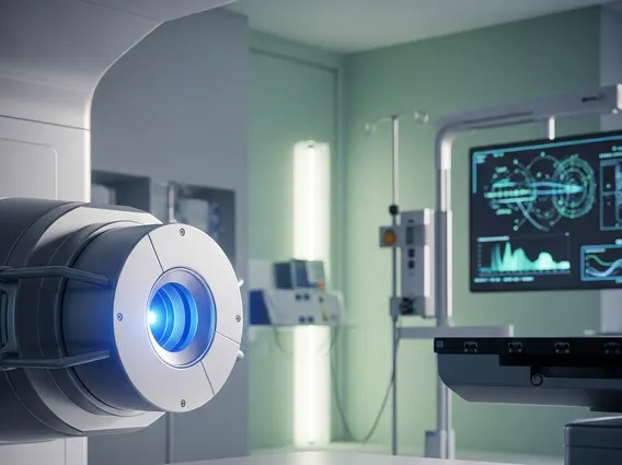
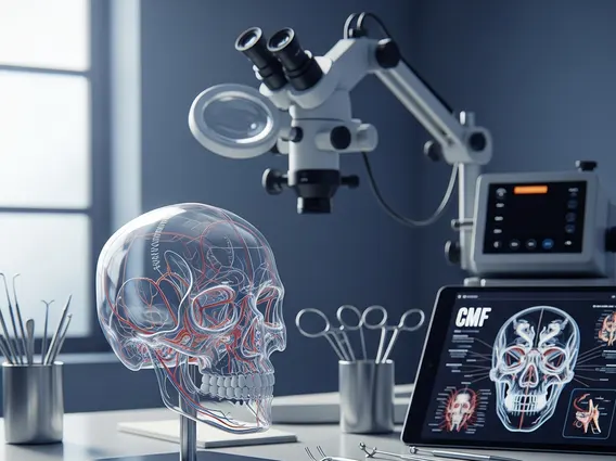
Fluorescence Guided Surgery refers to a cutting-edge surgical methodology that utilizes fluorescent dyes and specialized imaging systems to provide real-time visual guidance to surgeons. This technique allows for enhanced visualization of specific tissues, such as malignant tumors, lymph nodes, or blood vessels, that might otherwise be difficult to distinguish from surrounding healthy structures with the naked eye. By making these target tissues “glow,” surgeons can operate with greater precision, aiming to maximize the removal of diseased tissue while preserving healthy anatomy. The principle behind this approach involves administering a fluorescent agent that selectively accumulates in or binds to the target tissue.
Mechanism of Fluorescence Guided Surgery
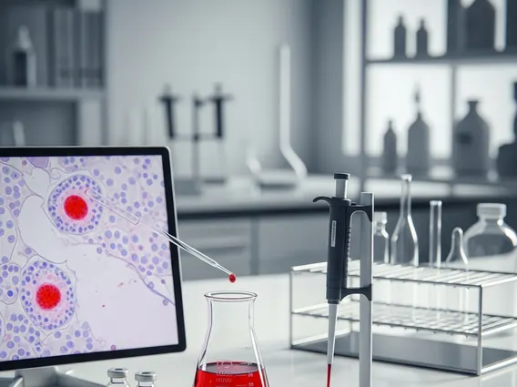
The mechanism of Fluorescence Guided Surgery involves several key steps, beginning with the administration of a fluorescent contrast agent to the patient. These agents are designed to selectively accumulate in or bind to specific cells or tissues, such as cancer cells, due to their unique biological properties or metabolic activity. Common agents include indocyanine green (ICG) or 5-aminolevulinic acid (5-ALA), which are activated by specific light wavelengths. Once the agent has circulated and localized in the target tissue, a specialized near-infrared (NIR) or visible light camera system is used during surgery. This system emits light at the excitation wavelength of the fluorescent agent, causing the agent within the target tissue to emit light at a different, longer wavelength (fluorescence). The camera then captures this emitted fluorescent signal, converting it into a real-time, high-contrast image that is displayed on a monitor in the operating room. This allows the surgical team to clearly visualize the target structures, such as tumor margins, blood flow, or nerve pathways, which are otherwise imperceptible.
- Administration of Fluorescent Agent: A specific dye is given to the patient, often intravenously, which then travels through the bloodstream.
- Selective Accumulation: The agent accumulates preferentially in target tissues (e.g., tumors) due to their distinct physiological characteristics.
- Light Excitation: During surgery, a specialized camera system emits light at a specific wavelength, exciting the fluorescent agent.
- Fluorescence Emission: The excited agent in the target tissue emits light at a different, detectable wavelength.
- Real-time Visualization: The camera captures this emitted light, displaying a high-contrast image on a screen, highlighting the target tissue for the surgeon.
Clinical Applications and Patient Benefits
Fluorescence Guided Surgery applications are diverse and rapidly expanding across numerous surgical disciplines, significantly enhancing surgical outcomes. In oncology, it is widely used for tumor resection in various cancers, including brain tumors, breast cancer, lung cancer, and colorectal cancer, enabling surgeons to achieve more complete removal of malignant tissue. For instance, in neurosurgery, 5-ALA is used to highlight glioblastoma cells, improving the extent of resection. Beyond cancer, it finds utility in cardiovascular surgery for assessing tissue perfusion and coronary artery bypass graft patency, and in lymphatic mapping for sentinel lymph node biopsies.
The benefits of Fluorescence Guided Surgery are substantial for both surgeons and patients. This technique offers enhanced precision, allowing surgeons to more accurately identify and remove diseased tissue while minimizing damage to surrounding healthy structures. This precision can lead to more complete resections, which is a critical factor in improving long-term patient survival rates, particularly in cancer surgery. Furthermore, it can reduce the incidence of positive surgical margins, potentially decreasing the need for subsequent surgeries or adjuvant therapies. Patients often experience reduced intraoperative blood loss, shorter operating times, and a lower risk of complications, contributing to faster recovery and improved quality of life. The ability to visualize critical structures like nerves and blood vessels also helps prevent iatrogenic injury, further safeguarding patient well-being.