Fine Needle Aspiration Biopsy
Fine Needle Aspiration Biopsy is a common diagnostic procedure used to investigate lumps or masses in various parts of the body. It offers a minimally invasive way to collect cells for examination, aiding in the diagnosis of conditions ranging from benign cysts to malignant tumors.

Key Takeaways
- Fine Needle Aspiration Biopsy (FNA) is a quick, minimally invasive procedure to collect cells for diagnosis.
- It involves using a thin needle to extract cell samples from suspicious lumps or masses.
- FNA is often guided by imaging techniques like ultrasound or CT for precision.
- Benefits include minimal discomfort, quick results, and avoidance of more invasive surgery.
- Results help differentiate between benign and malignant conditions, guiding further treatment decisions.
What is Fine Needle Aspiration Biopsy (FNA)?
Fine Needle Aspiration Biopsy (FNA) is a diagnostic technique that involves using a very thin, hollow needle to extract a small sample of cells from a suspicious lump or mass. This procedure is performed to determine the nature of the cells, specifically whether they are benign (non-cancerous) or malignant (cancerous). It is a widely used method for evaluating abnormalities found in organs such as the thyroid, breast, lymph nodes, and salivary glands, as well as soft tissue masses.
The primary goal of an FNA is to obtain cellular material for cytological examination by a pathologist. This examination helps in making an accurate diagnosis without the need for more invasive surgical procedures. The technique is particularly valuable for its speed, relative ease of performance, and minimal discomfort for the patient, providing crucial information that guides subsequent medical management and treatment planning.
Fine Needle Aspiration Biopsy Procedure
The fine needle aspiration biopsy procedure typically begins with the patient positioned comfortably. The area of interest is cleaned with an antiseptic solution, and a local anesthetic may be administered to numb the skin, though it’s often not required due to the needle’s thinness. The physician then carefully inserts a fine needle, often thinner than those used for blood draws, into the lump or mass.
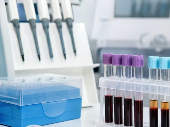
To ensure accuracy, the procedure is frequently performed under imaging guidance, such as ultrasound or computed tomography (CT) scans, especially for deeper or smaller lesions. Once the needle is in the correct position, the physician applies suction using a syringe to draw out cells and fluid. This process may be repeated several times to collect an adequate sample from different parts of the lesion. After the sample is collected, the needle is withdrawn, and pressure is applied to the site to minimize bruising. The collected cells are then smeared onto glass slides, fixed, and sent to a pathology laboratory for microscopic analysis.
Understanding FNA Biopsy Risks, Benefits, and Results
Understanding the FNA biopsy risks and benefits is crucial for patients considering this diagnostic tool. The benefits of FNA are significant, primarily its minimally invasive nature, which means less pain, a lower risk of complications compared to surgical biopsies, and a quicker recovery time. It is also a relatively fast procedure, often completed within 15-30 minutes, and typically allows for outpatient care. Furthermore, FNA can provide rapid preliminary results, helping clinicians make timely decisions about further investigations or treatment.
While generally safe, potential risks include minor bruising, swelling, or tenderness at the biopsy site. In rare cases, there might be slight bleeding or infection. Another consideration is the possibility of an inadequate sample, which might necessitate a repeat procedure. The fine needle aspiration biopsy results explained by a pathologist will categorize the findings, often as benign, malignant, suspicious for malignancy, atypical, or non-diagnostic. A benign result indicates no cancer, while a malignant result confirms cancer. Suspicious or atypical results may require further investigation, such as a core needle biopsy or surgical excision, to obtain a definitive diagnosis. Non-diagnostic results mean insufficient cells were collected, and the biopsy may need to be repeated.