Extramedullary Myeloid Tumor
An Extramedullary Myeloid Tumor, also known as a granulocytic sarcoma or chloroma, is a rare manifestation of myeloid neoplasms, characterized by the growth of immature myeloid cells outside the bone marrow. These tumors can appear in various parts of the body, presenting a diagnostic and therapeutic challenge.

Key Takeaways
- An Extramedullary Myeloid Tumor is a solid collection of immature myeloid cells occurring outside the bone marrow, often associated with acute myeloid leukemia (AML) or myelodysplastic syndromes (MDS).
- Symptoms vary widely based on the tumor’s location, ranging from skin lesions to neurological deficits or organ dysfunction.
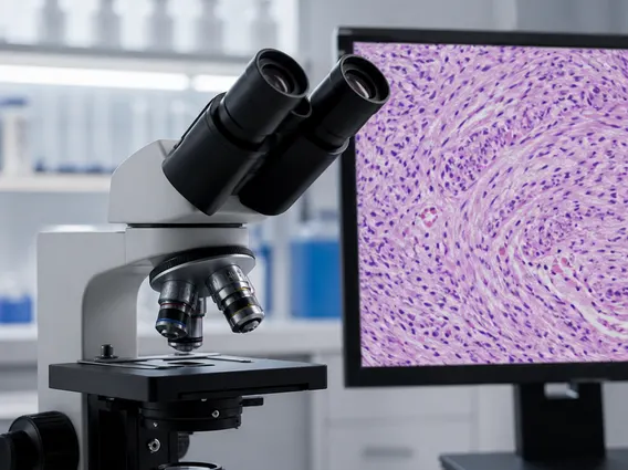
- Diagnosis typically involves biopsy of the affected tissue, followed by histopathological examination, immunohistochemistry, and molecular testing.
- Treatment strategies are highly individualized, often including chemotherapy, radiation therapy, surgery, and sometimes stem cell transplantation, depending on the underlying condition and tumor characteristics.
- Early and accurate diagnosis is crucial for effective management and improving patient outcomes.
What is an Extramedullary Myeloid Tumor?
An Extramedullary Myeloid Tumor refers to a localized collection of myeloid blast cells or immature myeloid cells that develops outside the bone marrow. These tumors are often associated with underlying myeloid malignancies such as acute myeloid leukemia (AML), myelodysplastic syndromes (MDS), or chronic myeloid leukemia (CML), but can also occur as the first sign of these diseases. Historically, they were known as “chloromas” due to their greenish appearance caused by myeloperoxidase, though not all tumors exhibit this color.
The presence of these tumors indicates that the cancerous myeloid cells have migrated from their usual site of production (the bone marrow) to other tissues. While relatively rare, their occurrence can signal disease progression or relapse. The specific characteristics of these tumors, including their cellular makeup and genetic profile, often mirror those of the underlying myeloid neoplasm, guiding both diagnosis and treatment approaches.
Recognizing Extramedullary Myeloid Tumor: Symptoms and Diagnosis
The presentation of an extramedullary myeloid tumor symptoms is highly variable, depending entirely on the tumor’s location and size. These tumors can affect virtually any organ system, leading to a diverse range of clinical manifestations. Common sites include the skin, lymph nodes, central nervous system (CNS), orbit, gastrointestinal tract, and soft tissues. For instance, skin involvement might present as firm, reddish-blue nodules, while CNS involvement could lead to neurological deficits such as weakness, sensory changes, or seizures. Tumors in the orbit may cause proptosis (bulging of the eye) or vision changes.
Accurate extramedullary myeloid tumor diagnosis is critical and typically involves a combination of clinical evaluation, imaging studies, and tissue biopsy. When a suspicious mass is identified, a biopsy is performed to obtain tissue for pathological examination. This tissue is then analyzed using:
- Histopathology: To identify the presence of immature myeloid cells and assess tissue architecture.
- Immunohistochemistry: To confirm the myeloid lineage of the cells by detecting specific markers (e.g., CD33, CD34, CD117, myeloperoxidase).
- Flow Cytometry: Can be used on fresh tissue samples to further characterize the cell population and identify aberrant phenotypes.
- Cytogenetics and Molecular Studies: To identify specific chromosomal abnormalities or gene mutations, which are crucial for classifying the underlying myeloid neoplasm and guiding prognosis and treatment.
Imaging techniques such as computed tomography (CT), magnetic resonance imaging (MRI), and positron emission tomography (PET) scans are often used to locate tumors, assess their extent, and monitor response to treatment. A bone marrow biopsy is also typically performed to evaluate for an underlying systemic myeloid malignancy.
Extramedullary Myeloid Tumor Treatment Options
The approach to extramedullary myeloid tumor treatment is highly individualized, taking into account the underlying myeloid neoplasm, the tumor’s location, size, and the patient’s overall health status. The primary goal is to eradicate the tumor and manage the associated systemic disease. Treatment options often include:
| Treatment Modality | Description | Considerations |
|---|---|---|
| Chemotherapy | Systemic chemotherapy, often similar to regimens used for acute myeloid leukemia, is a cornerstone of treatment. It targets rapidly dividing cancer cells throughout the body. | Effectiveness depends on the sensitivity of the tumor cells and the underlying disease. |
| Radiation Therapy | Localized radiation can be highly effective for controlling tumor growth, especially in sites where surgery is not feasible or for symptomatic relief. | Often used in conjunction with systemic therapy; dose and field are carefully planned to minimize side effects. |
| Surgery | Surgical removal may be considered for accessible tumors, particularly if they are causing significant symptoms or if diagnosis requires excisional biopsy. | Rarely curative on its own; typically followed by systemic therapy. |
| Targeted Therapy | Newer agents that specifically target molecular abnormalities found in myeloid neoplasms may be used, especially if the tumor cells express these targets. | Requires molecular testing of the tumor to identify suitable targets. |
| Hematopoietic Stem Cell Transplantation | For eligible patients with an underlying high-risk myeloid malignancy, stem cell transplantation may be considered as a consolidative therapy to prevent relapse. | A complex procedure with significant risks, reserved for specific patient populations. |
The choice of therapy is guided by the specific diagnosis of the myeloid neoplasm (e.g., AML, MDS, CML), the extent of extramedullary involvement, and the patient’s overall prognosis. Close monitoring is essential to assess treatment response and detect any recurrence. Multidisciplinary teams, including oncologists, radiation oncologists, surgeons, and pathologists, often collaborate to develop the most appropriate treatment plan for each patient.