Eus Fna
Eus Fna, or Endoscopic Ultrasound-Guided Fine Needle Aspiration, is a sophisticated medical procedure used to diagnose and stage various conditions, particularly those affecting the gastrointestinal tract and surrounding organs. This minimally invasive technique combines endoscopic imaging with targeted tissue sampling to provide crucial diagnostic information.

Key Takeaways
- EUS-FNA is a minimally invasive procedure combining endoscopy and ultrasound for precise tissue sampling.
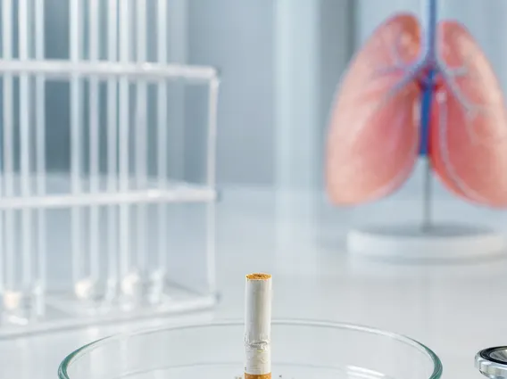
- It is primarily used for diagnosing and staging conditions in the gastrointestinal tract, pancreas, bile ducts, and mediastinum.
- The procedure involves inserting an endoscope with an ultrasound probe to visualize abnormalities and guide a fine needle for biopsy.
- Results from EUS-FNA are crucial for determining treatment plans, especially in oncology.
- While generally safe, potential risks include bleeding, infection, and pancreatitis, though serious complications are rare.
What is Eus Fna?
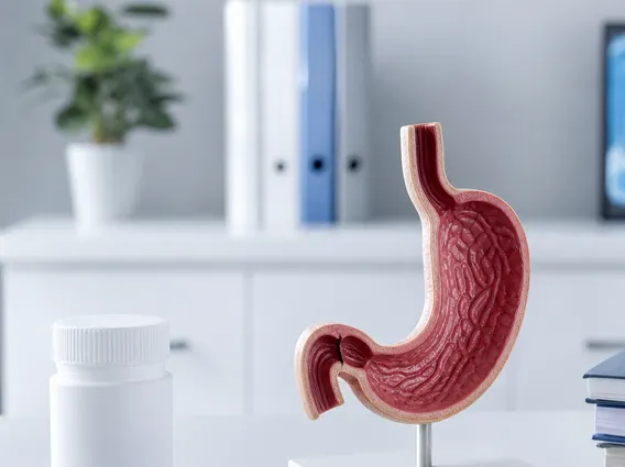
Eus Fna, or Endoscopic Ultrasound-Guided Fine Needle Aspiration, is a diagnostic procedure that merges two advanced medical technologies: endoscopy and endoscopic ultrasound (EUS). This technique allows physicians to visualize and obtain tissue samples (biopsies) from lesions or abnormalities located deep within the body, often inaccessible by conventional methods. The procedure provides a comprehensive Eus Fna definition and explanation by allowing direct visualization of internal organs and structures, such as the pancreas, bile ducts, lymph nodes, and lesions in the gastrointestinal wall, followed by precise tissue acquisition.
The primary goal of EUS-FNA is to accurately diagnose various conditions, including cancers, benign tumors, cysts, and inflammatory processes. By obtaining cellular material or tissue, pathologists can analyze the samples to determine the nature of the abnormality. This detailed analysis is vital for guiding treatment decisions, especially in oncology, where accurate staging and characterization of tumors significantly impact patient management. To learn about Eus Fna involves understanding its dual imaging and sampling capabilities, which make it an invaluable tool in modern gastroenterology and pulmonology.
The Eus Fna Procedure: Indications and Process
The EUS-FNA procedure is indicated for a wide range of diagnostic purposes. Common indications include the evaluation of pancreatic masses, staging of esophageal, gastric, and rectal cancers, assessment of mediastinal lymphadenopathy (enlarged lymph nodes in the chest), and characterization of submucosal lesions in the gastrointestinal tract. It offers a detailed Eus Fna overview of internal structures that might be obscured by gas or bone in other imaging modalities, providing high-resolution images.
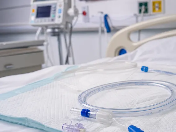
The process typically begins with the patient receiving conscious sedation or general anesthesia. An endoscope equipped with a miniature ultrasound transducer at its tip is then gently guided through the mouth (for upper GI and mediastinal access) or rectum (for lower GI access). Once the target lesion or abnormality is identified using the ultrasound images, a very fine needle is passed through a channel in the endoscope. Under real-time ultrasound guidance, the needle is advanced into the lesion, and a small sample of cells or tissue is aspirated. This precise targeting minimizes damage to surrounding healthy tissue and maximizes the diagnostic yield. Multiple passes may be performed to ensure an adequate sample is obtained. The collected samples are then sent to a pathology laboratory for microscopic examination.
Key steps in the EUS-FNA procedure include:
- Patient preparation (fasting, sedation).
- Insertion of the echoendoscope.
- Ultrasound visualization and localization of the target lesion.
- Passage of the fine needle through the endoscope.
- Aspiration of tissue or cells under real-time ultrasound guidance.
- Withdrawal of the needle and endoscope.
- Preparation of samples for pathological analysis.
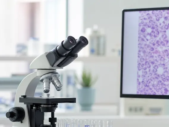
Interpreting Eus Fna Results and Potential Risks
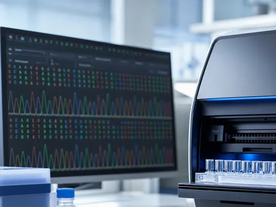
Interpreting EUS-FNA results is a critical step in patient care. Once the tissue samples are analyzed by a pathologist, a detailed report is generated. This report will classify the nature of the cells, indicating whether they are benign (non-cancerous), malignant (cancerous), or indeterminate. For malignant findings, the report may also provide information on the type of cancer, its grade, and sometimes specific molecular markers, which are vital for personalized treatment strategies. The accuracy of EUS-FNA in diagnosing pancreatic masses, for instance, is reported to be high, often exceeding 85-90%, making it a cornerstone in the management of such conditions.
While EUS-FNA is generally considered a safe procedure, like any invasive medical intervention, it carries potential risks. These risks are typically low but can include bleeding at the biopsy site, infection (especially if a cyst is punctured), and pancreatitis (inflammation of the pancreas), particularly when sampling pancreatic lesions. Other less common risks include perforation of the gastrointestinal tract or adverse reactions to sedation. Patients are usually monitored for a few hours post-procedure and advised on potential symptoms to watch for. It is crucial for patients to discuss any concerns with their healthcare provider to understand the benefits and risks specific to their situation.