
Myelogram
A myelogram is a specialized diagnostic imaging procedure used to examine the spinal canal, spinal cord, nerve roots, and other surrounding structures. It involves injecting a contrast dye into the cerebrospinal fluid (CSF) space before taking X-rays or a CT scan.
Key Takeaways
- A myelogram is an imaging test that uses contrast dye to visualize the spinal canal and its contents.
- It helps diagnose conditions like herniated discs, spinal stenosis, and tumors affecting the spinal cord or nerve roots.
- The procedure involves a lumbar puncture to inject contrast, followed by X-rays or a CT scan.
- Common risks include headache and nausea, with rare but more serious complications possible.
- Recovery typically involves rest, hydration, and monitoring for post-procedure symptoms.
What is Myelogram: Definition and Purpose
A Myelogram is a diagnostic imaging test that uses a special contrast material and X-rays or computed tomography (CT) to look for problems in the spinal canal. This procedure specifically visualizes the spinal cord, nerve roots, and the subarachnoid space where cerebrospinal fluid (CSF) circulates. The contrast dye highlights these structures, allowing radiologists to identify abnormalities that might not be visible on standard X-rays or even some MRI scans.
The primary purpose of a myelogram is to diagnose conditions affecting the spinal cord and nerve roots. It is particularly useful when other imaging techniques, such as magnetic resonance imaging (MRI), are inconclusive or contraindicated (e.g., due to pacemakers or certain metallic implants). Conditions commonly identified or evaluated with a myelogram include:
- Herniated or bulging discs compressing nerves.
- Spinal stenosis (narrowing of the spinal canal).
- Spinal tumors or cysts.
- Inflammation or infection affecting the spinal cord or nerve roots.
- Bone spurs (osteophytes) causing nerve impingement.
By providing detailed images of the spinal canal, a myelogram helps clinicians pinpoint the exact location and nature of spinal issues, guiding appropriate treatment plans, which may include surgery or other interventions.
The Myelogram Procedure Explained
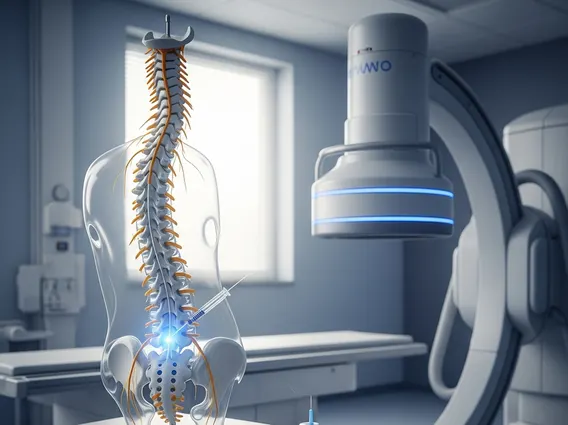
The myelogram procedure explained involves several key steps, typically performed in a hospital or outpatient imaging center by a radiologist. Before the procedure, patients are usually advised to fast for a few hours and may need to adjust certain medications, especially blood thinners. Upon arrival, the patient lies on an X-ray table, and the injection site, usually in the lower back (lumbar region), is cleaned and numbed with a local anesthetic.
The radiologist then performs a lumbar puncture, carefully inserting a thin needle into the subarachnoid space to access the cerebrospinal fluid. A small amount of CSF may be collected for analysis, and then the contrast dye is slowly injected. Fluoroscopy, a type of real-time X-ray, is used to guide the needle and monitor the flow of the contrast material as it coats the spinal cord and nerve roots. After the injection, the patient may be tilted on the table to allow the contrast to flow to different areas of the spine, depending on the region being examined.
Once the contrast has adequately distributed, a series of X-ray images are taken. In many cases, a computed tomography (CT) scan immediately follows the myelogram to provide even more detailed cross-sectional images of the spine. The entire procedure typically takes about 30 to 60 minutes, though preparation and post-procedure observation can extend the total time. After the imaging is complete, the needle is removed, and a bandage is applied to the injection site.
Myelogram Risks and Recovery
Understanding myelogram risks and recovery is crucial for patients undergoing this diagnostic test. While generally safe, like any invasive procedure, a myelogram carries potential risks. The most common side effect is a headache, often referred to as a “post-dural puncture headache,” which can range from mild to severe and may last for several days. This occurs due to a small leakage of cerebrospinal fluid from the puncture site. Other common, usually mild, side effects include nausea, dizziness, vomiting, and temporary neck or back stiffness.
Less common but more serious risks include allergic reactions to the contrast dye, infection (meningitis), bleeding at the puncture site, nerve damage, or, very rarely, seizures. Patients are carefully screened for allergies and medical conditions that might increase these risks before the procedure. Following the myelogram, patients are typically advised to lie flat for several hours to minimize the risk of headache and to drink plenty of fluids to help replenish CSF. Activity restrictions, such as avoiding strenuous exercise or heavy lifting, are usually recommended for 24 to 48 hours.
Most individuals experience a full recovery within a few days. It is important for patients to monitor for any concerning symptoms after the procedure, such as severe or persistent headache not relieved by pain medication, fever, stiff neck, numbness or weakness in the legs, or any signs of infection at the injection site. If any of these symptoms occur, immediate medical attention should be sought.


