Squamous Cell Carcinoma Signs & Symptoms
Squamous cell carcinoma (SCC) is a common form of skin cancer that arises from the squamous cells in the outer layer of the skin. Recognizing the squamous cell carcinoma symptoms and early warning signs is crucial for timely diagnosis and effective treatment.
Key Takeaways
- Squamous cell carcinoma symptoms often appear as persistent red patches, scaly growths, or non-healing sores on sun-exposed skin.
- Early signs of squamous cell carcinoma include new or changing skin lesions that may be tender or bleed easily.
- Knowing what does squamous cell carcinoma look like—such as raised, firm nodules or open ulcers—is vital for self-examination.
- Regular self-skin exams and professional dermatological checks are essential for identifying squamous cell carcinoma warning signs.
- Prompt medical evaluation of any suspicious skin changes significantly improves treatment outcomes for SCC.
Understanding Squamous Cell Carcinoma Symptoms
Understanding the common squamous cell carcinoma symptoms is the first step in early detection. These symptoms often manifest on areas of the skin frequently exposed to the sun, such as the face, ears, neck, lips, and hands. Unlike some other skin conditions, SCC lesions tend to persist and may even grow over time, indicating a need for medical attention. According to the American Academy of Dermatology, SCC is the second most common type of skin cancer, with over 1 million cases diagnosed annually in the United States, highlighting the importance of public awareness regarding its signs.
Persistent Red Patches
One of the initial symptoms of SCC skin cancer can be the appearance of persistent red patches on the skin. These patches might be flat or slightly raised and can sometimes be mistaken for eczema, psoriasis, or a common rash. However, unlike benign skin irritations, these patches do not typically respond to over-the-counter creams or home remedies and tend to linger for weeks or months. They may also feel rough or scaly to the touch, distinguishing them from simple redness.
Scaly, Crusted Growths
Another common presentation of squamous cell carcinoma symptoms involves scaly or crusted growths. These lesions often have a rough, sandpaper-like texture and may be white, pink, or red. They can develop a crust or scab that might bleed if scratched or bumped, and then reappear. These growths can be tender or painful, especially if they are located in areas prone to friction or pressure. The persistent nature of these scaly, crusted areas, particularly in sun-damaged skin, should raise suspicion.
Recognizing Early Signs of SCC
Recognizing the early signs of squamous cell carcinoma is critical for improving prognosis. Early detection allows for less invasive treatment options and a higher cure rate. Many people might overlook subtle changes, attributing them to aging or minor skin issues, but vigilance is key. Learning how to identify squamous cell carcinoma in its nascent stages can make a significant difference in health outcomes.
New or Changing Skin Lesions
Any new skin lesion that appears suddenly and persists, or an existing lesion that begins to change in size, shape, or color, should be considered an early sign of squamous cell carcinoma. These changes can be subtle, such as a slight increase in diameter, a change in texture, or the development of new symptoms like itching or tenderness. Regular self-skin examinations are crucial for monitoring these changes, particularly on areas of the body that are frequently exposed to the sun.
Non-Healing Wounds
A wound, sore, or ulcer that does not heal within a few weeks is a significant squamous cell carcinoma warning sign. Unlike typical cuts or scrapes that heal relatively quickly, an SCC-related lesion might remain open, bleed periodically, and fail to form a stable scab. These non-healing areas can sometimes be mistaken for insect bites or persistent acne, but their prolonged presence and lack of improvement warrant immediate medical evaluation. This is a key indicator when trying to identify signs of squamous cell carcinoma on skin.
What Squamous Cell Carcinoma Looks Like
Understanding what does squamous cell carcinoma look like is essential for both self-detection and aiding medical professionals in diagnosis. While its appearance can vary, there are several common visual characteristics that can help in identifying this type of skin cancer. These visual cues are often distinct from benign moles or other skin conditions, making it possible for individuals to notice potential issues during routine self-checks.
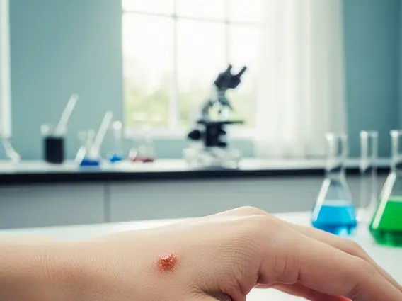
Raised, Firm Nodules
One common presentation of SCC is a raised, firm nodule. These growths can vary in color from flesh-toned to reddish-brown and often have a hard, indurated feel. They may grow relatively quickly and can sometimes develop a central depression or ulceration. These nodules are often found on areas of chronic sun exposure and can be tender to the touch. The presence of such a firm, growing lump is a strong indicator of squamous cell carcinoma on skin.
Open Sores or Ulcers
In more advanced stages, or sometimes from the outset, SCC can present as an open sore or ulcer. These lesions may have an irregular border and a crusted or bleeding surface. They often fail to heal, or they might heal partially and then break open again. The base of the ulcer may appear red or inflamed. This type of lesion can be particularly concerning because it indicates a breakdown of the skin barrier and potential for deeper tissue involvement, making it a critical squamous cell carcinoma warning sign.
Identifying High-Risk Areas and Warning Signs
Identifying squamous cell carcinoma warning signs also involves understanding where these lesions are most likely to appear and what specific characteristics make them suspicious. Awareness of high-risk areas and specific lesion features can significantly improve the chances of early detection and successful treatment. Knowing how to identify squamous cell carcinoma effectively involves a combination of visual recognition and an understanding of risk factors.
Sun-Exposed Body Parts
The vast majority of SCCs develop on areas of the body that receive chronic and intense sun exposure. These include the face, especially the nose, lips, ears, and forehead, as well as the scalp (particularly in bald individuals), neck, hands, and forearms. Less commonly, SCC can appear on other parts of the body, including mucous membranes or areas of chronic inflammation or scarring. The cumulative effect of ultraviolet (UV) radiation from the sun is the primary cause, making these areas particularly susceptible to signs of squamous cell carcinoma on skin.
Lesions with Irregular Borders
While not as commonly associated with SCC as with melanoma, some SCC lesions can exhibit irregular or ill-defined borders. Unlike benign growths that often have smooth, symmetrical edges, an SCC may have an uneven or notched perimeter. This irregularity, combined with other features like persistent scaling, crusting, or non-healing, makes it a significant squamous cell carcinoma warning sign. Any lesion that changes in shape or develops an irregular border should be promptly evaluated by a dermatologist to determine if it is among the squamous cell carcinoma symptoms requiring attention.
Frequently Asked Questions
What are the primary risk factors for Squamous Cell Carcinoma?
The primary risk factor for SCC is prolonged exposure to ultraviolet (UV) radiation from sunlight or tanning beds. Other significant factors include fair skin, a history of sunburns, older age, a weakened immune system (e.g., organ transplant recipients), certain genetic syndromes, and previous radiation therapy. Individuals with a history of actinic keratoses, which are precancerous lesions, are also at higher risk. Understanding these factors helps in assessing personal risk and implementing preventive measures.
How is Squamous Cell Carcinoma diagnosed?
Diagnosis of SCC typically begins with a visual examination by a dermatologist. If a suspicious lesion is identified, a biopsy is performed. This involves removing a small tissue sample from the lesion, which is then examined under a microscope by a pathologist. The biopsy confirms the presence of cancer cells and determines the type of skin cancer. Early and accurate diagnosis through biopsy is crucial for guiding appropriate treatment decisions.
Is Squamous Cell Carcinoma curable?
Yes, squamous cell carcinoma is highly curable, especially when detected early and treated promptly. The cure rate is very high, often exceeding 95%, for lesions identified and treated in their early stages. Treatment options typically include surgical removal (such as Mohs surgery or excisional surgery), radiation therapy, or cryosurgery. Regular follow-up examinations are recommended to monitor for recurrence or the development of new lesions.