
Stage Iia Intraocular Melanoma Of The Ciliary Body And Choroid
Stage IIa intraocular melanoma of the ciliary body and choroid represents a specific classification of a rare eye cancer. This article provides an overview of this condition, its symptoms, and the available treatment options and prognosis.
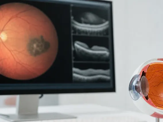
Key Takeaways
- Stage IIa Intraocular Melanoma of the Ciliary Body and Choroid is a rare eye cancer affecting the middle layers of the eye.
- Early stages may present with subtle or no symptoms, making regular eye exams crucial for detection.
- Common symptoms can include blurred vision, flashes of light, floaters, or a dark spot in the vision.
- Treatment options vary but often involve radiation therapy or surgical removal, depending on tumor characteristics.
- The prognosis for stage IIa intraocular melanoma is generally favorable compared to more advanced stages, especially with timely intervention.
What is Stage IIa Intraocular Melanoma of the Ciliary Body and Choroid?
Stage IIa Intraocular Melanoma of the Ciliary Body and Choroid refers to a specific classification of a malignant tumor originating in the uvea, the middle layer of the eye. Intraocular melanoma is the most common primary cancer inside the eye in adults, though it is still considered rare, with an incidence of about 5-6 cases per million people per year in the United States, according to the American Cancer Society. This particular stage, Stage IIa, indicates a tumor that has reached a certain size (typically between 3.1 to 8.0 mm in thickness and up to 16 mm in greatest basal diameter) but has not spread beyond the eye. The ciliary body and choroid are parts of the uvea; the choroid is a vascular layer that provides oxygen and nutrients to the retina, while the ciliary body produces aqueous humor and contains muscles that change the shape of the lens for focusing.
Understanding what is stage IIa intraocular melanoma involves recognizing its location and size criteria. Tumors in these specific regions can sometimes be more challenging to detect early due to their position within the eye, potentially growing larger before symptoms become noticeable.
Symptoms of Stage IIa Intraocular Melanoma
The symptoms of ciliary body and choroid melanoma can vary depending on the tumor’s size and exact location within the eye. In its early stages, Stage IIa intraocular melanoma may not cause any noticeable symptoms, often being discovered during a routine eye examination. As the tumor grows, it can begin to affect vision or cause other ocular disturbances.
Common symptoms that may prompt a visit to an ophthalmologist include:
- Blurred vision or a decrease in visual acuity.
- Seeing flashes of light (photopsia) or new floaters (small specks or strings that drift across the field of vision).
- A dark spot in the field of vision or a blind spot.
- Changes in the shape of the pupil.
- Eye pain, though this is less common and usually indicates a larger or more advanced tumor.
- A change in the position of the eyeball within the socket (proptosis) or pressure behind the eye.
It is important to note that many of these symptoms can also be caused by less serious eye conditions. However, persistent or worsening symptoms warrant immediate medical evaluation by an eye care professional.
Treatment Options and Prognosis for Stage IIa Intraocular Melanoma
The approach to intraocular melanoma stage IIa treatment is highly individualized, depending on factors such as tumor size, location, the patient’s overall health, and the treating physician’s expertise. The primary goal of treatment is to destroy the tumor, preserve vision, and prevent the cancer from spreading (metastasis).
Common treatment modalities include:
- Radiation Therapy: This is a very common treatment for intraocular melanoma. It can involve brachytherapy (plaque radiotherapy), where a small radioactive disc is surgically placed on the eye over the tumor for several days, or external beam radiation therapy, such as proton beam therapy, which delivers highly focused radiation to the tumor.
- Surgical Removal: In some cases, particularly for smaller tumors or those in specific locations, surgical removal of the tumor (transscleral resection) may be an option. For larger tumors or those that have severely impacted vision, enucleation (surgical removal of the entire eye) might be recommended, followed by fitting a prosthetic eye.
- Laser Therapy: Transpupillary thermotherapy (TTT) uses a laser to heat and destroy small tumors, sometimes used in conjunction with radiation.
The prognosis for stage IIa intraocular melanoma is generally favorable, especially when detected and treated early. According to the American Cancer Society, the 5-year survival rate for localized ocular melanoma (which includes Stage IIa) is approximately 85%. However, prognosis can be influenced by several factors, including the tumor’s size, its location (tumors in the ciliary body may have a slightly less favorable prognosis than those in the choroid due to their proximity to drainage pathways), and certain genetic markers within the tumor. Regular follow-up examinations are crucial after treatment to monitor for recurrence and to detect any potential spread of the cancer to other parts of the body, though this is less common in Stage IIa.



















