Pulmonary Sulcus Tumor
A Pulmonary Sulcus Tumor is a specific type of lung cancer located in the apex of the lung, often presenting unique challenges due to its proximity to vital structures. Understanding this condition is crucial for early detection and effective management.
Key Takeaways
- Pulmonary Sulcus Tumor is a form of lung cancer situated at the lung’s apex, distinct from other lung tumors.
- Its location near nerves and blood vessels leads to characteristic symptoms, including shoulder and arm pain.
- Diagnosis involves imaging like CT and MRI, often followed by biopsy for confirmation.
- Treatment typically combines surgery, radiation, and sometimes chemotherapy, tailored to the individual case.
- Early recognition and a multidisciplinary approach are vital for improving patient outcomes.
What is a Pulmonary Sulcus Tumor?
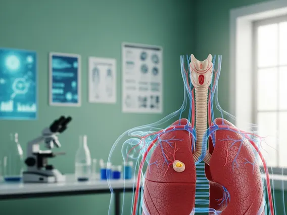
Pulmonary Sulcus Tumor refers to a relatively rare form of lung cancer that develops in the superior sulcus, the groove at the top of the lung. These tumors are often non-small cell lung cancers and are particularly challenging due to their anatomical location. They are situated close to critical structures such as the brachial plexus (a network of nerves controlling arm and hand movement), subclavian vessels, sympathetic nerves, and vertebrae. This proximity often leads to a distinct set of symptoms known as Pancoast syndrome. The incidence of lung cancer overall is significant, with an estimated 238,340 new cases in the United States in 2023, according to the American Cancer Society, though Pulmonary Sulcus Tumors represent a smaller subset of these cases.
Recognizing Pulmonary Sulcus Tumor: Symptoms and Diagnosis
Recognizing a Pulmonary Sulcus Tumor often begins with its characteristic symptoms, which are primarily due to the tumor’s invasion of surrounding structures. The most common presentation is severe, persistent pain in the shoulder, arm, and hand, often radiating along the ulnar nerve distribution. Other symptoms can include muscle weakness or atrophy in the hand, Horner’s syndrome (a combination of drooping eyelid, constricted pupil, and decreased sweating on one side of the face), and sometimes superior vena cava syndrome. These symptoms collectively are known as Pancoast syndrome.
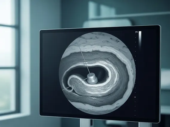
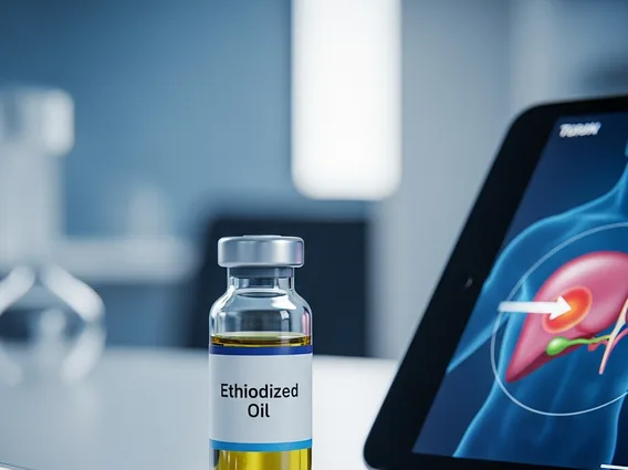
The Pulmonary sulcus tumor diagnosis process typically involves a combination of imaging studies and tissue biopsy. Initial evaluation may include a chest X-ray, but more detailed imaging is crucial for accurate staging and treatment planning:
- Computed Tomography (CT) scan: Provides detailed cross-sectional images of the chest, helping to define tumor size and local invasion.
- Magnetic Resonance Imaging (MRI) scan: Essential for assessing the extent of soft tissue involvement, particularly invasion of the brachial plexus, vertebral bodies, or spinal canal.
- Positron Emission Tomography (PET) scan: Used to detect distant metastases and evaluate metabolic activity of the tumor.
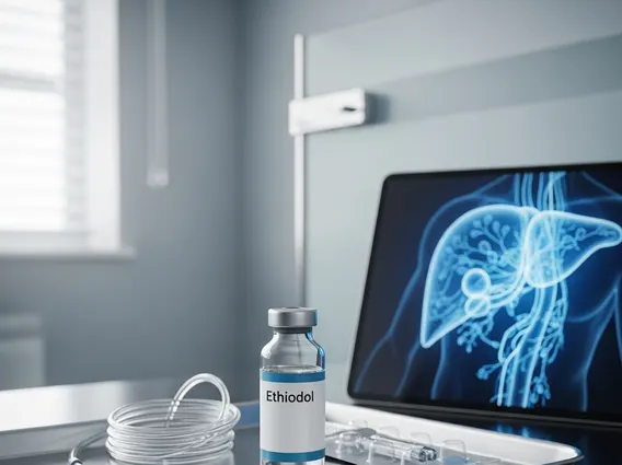
A definitive diagnosis requires a biopsy, which can be obtained via bronchoscopy, CT-guided needle biopsy, or surgical biopsy. Pathological examination confirms the type of cancer and guides further treatment decisions.
Treatment Approaches for Pulmonary Sulcus Tumors
The Pulmonary sulcus tumor treatment strategy is complex and highly individualized, often requiring a multidisciplinary approach involving thoracic surgeons, radiation oncologists, and medical oncologists. Due to the tumor’s challenging location and potential for local invasion, treatment typically involves a combination of modalities.
Historically, surgery was the primary treatment, but neoadjuvant therapy (treatment given before surgery) has become standard practice to improve resectability and outcomes. This often includes chemotherapy, administered to shrink the tumor and treat any microscopic spread, and radiation therapy, used to reduce tumor size and control local disease, often in conjunction with chemotherapy. External beam radiation therapy (EBRT) is commonly employed, sometimes with advanced techniques like intensity-modulated radiation therapy (IMRT) to spare healthy tissues.
Following neoadjuvant therapy, surgical resection is often performed if the tumor has responded well and is deemed resectable. Surgical approaches can be challenging and may involve removing parts of the ribs, vertebrae, or blood vessels depending on the extent of invasion. For patients who are not surgical candidates, definitive chemoradiation therapy may be the primary treatment. Post-operative radiation therapy may also be considered in some cases to reduce the risk of recurrence. Ongoing research continues to explore new therapeutic avenues, including targeted therapies and immunotherapy, to further improve outcomes for individuals with this challenging condition.