
Proton Magnetic Resonance Spectroscopic Imaging
Proton Magnetic Resonance Spectroscopic Imaging (PMSRI) is an advanced diagnostic technique that provides detailed biochemical information about tissues within the body. It complements traditional magnetic resonance imaging (MRI) by offering insights into metabolic changes associated with various diseases.
Key Takeaways
- Proton Magnetic Resonance Spectroscopic Imaging (PMSRI) is a non-invasive medical imaging technique that analyzes the chemical composition of tissues.
- It works by detecting signals from hydrogen protons in different molecules, revealing metabolic profiles.
- PMSRI provides crucial biochemical information beyond anatomical details, aiding in disease characterization.
- Key applications include differentiating tumor types, assessing treatment response, and diagnosing metabolic disorders in the brain.
- The technique helps clinicians make more informed decisions by offering a unique window into tissue biochemistry.
What is Proton Magnetic Resonance Spectroscopic Imaging?
Proton Magnetic Resonance Spectroscopic Imaging (PMSRI) is a sophisticated medical imaging modality that extends the capabilities of conventional magnetic resonance imaging (MRI). While MRI primarily provides anatomical details, PMSRI focuses on detecting and quantifying specific metabolites within tissues. This allows clinicians to gain a biochemical understanding of the body’s processes, which can be critical for diagnosing and monitoring various conditions. Essentially, proton magnetic resonance spectroscopy explained through PMSRI maps the distribution and concentration of hydrogen-containing compounds, such as lactate, choline, creatine, and N-acetylaspartate (NAA), which are vital indicators of cellular health and disease states.
Principles and Mechanism of Proton MR Spectroscopic Imaging
Understanding how proton MR spectroscopic imaging works involves grasping its foundation in nuclear magnetic resonance principles. PMSRI utilizes strong magnetic fields and radiofrequency pulses, similar to standard MRI, but with a crucial difference: it analyzes the subtle variations in the resonant frequency of hydrogen protons based on their chemical environment. Different molecules in the body contain hydrogen atoms, and the electrons surrounding these atoms slightly shield the protons from the main magnetic field. This shielding effect, known as the “chemical shift,” causes protons in different molecules to resonate at slightly different frequencies.
The process involves:
- Magnetic Field Application: The patient is placed in a powerful magnetic field, aligning the spins of hydrogen protons in the body’s water and metabolites.
- Radiofrequency Pulse: A brief radiofrequency pulse is applied, knocking the aligned protons out of alignment.
- Signal Detection: As the protons relax back to their aligned state, they emit radiofrequency signals.
- Chemical Shift Analysis: These emitted signals are then detected and analyzed. The slight differences in frequency (chemical shifts) reveal the specific molecules present and their concentrations.
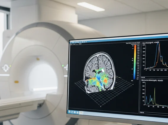
- Spatial Mapping: By acquiring signals from specific spatial locations (voxels), PMSRI creates a map of metabolite concentrations across the region of interest, providing both biochemical and spatial information.
This detailed analysis allows for the creation of a “metabolic fingerprint” of the tissue, which can be indicative of normal physiological function or pathological changes.
Clinical Applications of Proton MRS Imaging
The applications of proton MRS imaging are diverse and continually expanding, particularly in neurology and oncology. This technique offers unique insights that are often unattainable with conventional imaging alone. For instance, in neuro-oncology, PMSRI is invaluable for characterizing brain tumors. It can help differentiate between various tumor types, distinguish tumor recurrence from radiation necrosis, and assess the aggressiveness of a lesion. For example, high levels of choline and low levels of NAA are often indicative of malignancy.
Here are some key clinical applications:
- Brain Tumor Characterization: Aids in grading tumors, planning biopsies, and monitoring treatment response by detecting changes in metabolite ratios (e.g., elevated choline, reduced NAA).
- Stroke and Ischemia: Can identify areas of metabolic distress by detecting increased lactate, indicating anaerobic metabolism due to reduced blood flow.
- Neurodegenerative Diseases: Provides insights into conditions like Alzheimer’s disease and Parkinson’s disease by showing changes in NAA (neuronal marker) and myo-inositol (glial marker).
- Epilepsy: Helps localize seizure foci by identifying regions with altered metabolism.
- Infectious and Inflammatory Conditions: Can differentiate between various brain lesions, such as abscesses or demyelinating plaques, based on their unique metabolic profiles.
PMSRI serves as a powerful tool, complementing anatomical imaging to provide a more comprehensive understanding of disease processes, thereby guiding more precise diagnostic and therapeutic strategies.


