Papilledema
Papilledema is a serious medical condition characterized by the swelling of the optic disc, the part of the optic nerve visible inside the eye. This swelling is a critical sign of increased pressure within the skull, known as intracranial pressure.
Key Takeaways
- Papilledema indicates optic disc swelling due to elevated intracranial pressure.
- It is often asymptomatic early but can lead to visual disturbances and permanent vision loss if untreated.
- Common causes include brain tumors, hydrocephalus, and idiopathic intracranial hypertension.
- Diagnosis involves ophthalmoscopy, neuroimaging, and often a lumbar puncture.
- Treatment focuses on addressing the underlying cause of the increased intracranial pressure.
What is Papilledema?
What is Papilledema refers to the swelling of the optic disc, where the optic nerve enters the eyeball. This condition is not a disease itself but a clinical sign indicating elevated intracranial pressure (ICP) within the skull. The optic nerve, which transmits visual information, is directly affected by changes in cerebrospinal fluid (CSF) pressure. When ICP rises, it impedes venous return from the retina and causes optic nerve axons to swell at the disc.
While papilledema can affect individuals of any age, it is relatively uncommon. A study published in “Neurology” indicated that the incidence of idiopathic intracranial hypertension (a common cause of papilledema) is approximately 1-2 per 100,000 people per year, with higher rates observed in obese women of childbearing age. Early detection is crucial because sustained optic disc swelling can lead to irreversible optic nerve damage and permanent vision loss.
Symptoms and Diagnosis of Papilledema
The presentation of papilledema symptoms and causes can vary significantly. In early stages, it may be asymptomatic. As the condition progresses, symptoms often include transient visual obscurations (brief episodes of blurred or dimmed vision), headaches, nausea, vomiting, and pulsatile tinnitus (a whooshing sound in the ears synchronized with the heartbeat). Visual field defects, such as an enlarged blind spot, can also develop. Underlying causes are diverse, ranging from serious neurological conditions like brain tumors, hydrocephalus, and cerebral venous sinus thrombosis, to inflammatory conditions, severe hypertension, or certain medications. A common cause, particularly in young, obese women, is idiopathic intracranial hypertension (IIH), where ICP is elevated without an identifiable secondary cause.
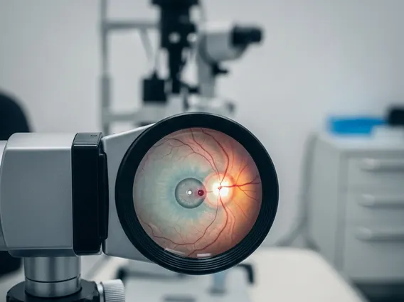
How is papilledema diagnosed typically involves a comprehensive eye examination, with ophthalmoscopy being key. During this, an ophthalmologist or neurologist observes the optic disc for signs of swelling, such as blurring of disc margins, elevation, and absence of the optic cup. Further diagnostic steps are essential to determine the underlying cause of increased ICP, often including:
- Neuroimaging: MRI of the brain and orbits, sometimes with venography (MRV), to rule out structural abnormalities like tumors or venous sinus thrombosis.
- Lumbar Puncture: If neuroimaging is normal, a spinal tap measures cerebrospinal fluid opening pressure and analyzes its composition, confirming elevated ICP and excluding inflammatory causes.
- Visual Field Testing: This assesses the extent of any peripheral vision loss, indicating optic nerve damage.
Managing Papilledema: Treatment Options
Effective papilledema treatment options are primarily directed at reducing elevated intracranial pressure and addressing its underlying cause. Since papilledema is a sign rather than a primary disease, treating the root cause is paramount to preserving vision and alleviating symptoms.
Treatment strategies vary widely depending on the specific etiology:
- For brain tumors: Surgical removal, radiation therapy, or chemotherapy may be necessary to reduce tumor size and subsequent ICP.
- For hydrocephalus: A shunt procedure (e.g., ventriculoperitoneal shunt) might be implanted to drain excess CSF, lowering pressure.
- For idiopathic intracranial hypertension (IIH): Weight loss is often recommended for obese patients. Medications like acetazolamide decrease CSF production. In severe cases, optic nerve sheath fenestration or CSF shunting may be considered.
- For cerebral venous sinus thrombosis: Anticoagulant medications are used to dissolve blood clots.
- For severe hypertension: Aggressive management of blood pressure is crucial to reduce ICP.
Regular follow-up with an ophthalmologist and neurologist is essential to monitor optic nerve health, visual function, and ICP levels, ensuring the treatment plan is effective and adjusted as needed.