Papillary Intralymphatic Angioendothelioma
Papillary Intralymphatic Angioendothelioma (PILA) is a rare and distinct vascular tumor characterized by its unique growth pattern within lymphatic vessels. This condition, though generally considered benign, exhibits local aggressiveness and requires careful diagnosis and management.
Key Takeaways
- Papillary Intralymphatic Angioendothelioma is a rare, benign vascular tumor primarily affecting lymphatic vessels.
- It typically presents as a slow-growing mass, often accompanied by pain or swelling, and can occur in various parts of the body.
- The exact Papillary Intralymphatic Angioendothelioma causes are not fully understood, but it is not considered hereditary.
- Diagnosis relies on histopathological examination of a biopsy, often supported by imaging studies.
- Treatment primarily involves surgical removal, with long-term follow-up essential due to the potential for local recurrence.
What is Papillary Intralymphatic Angioendothelioma?
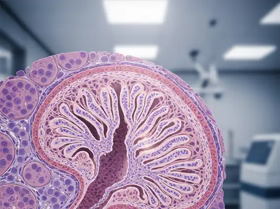
What is Papillary Intralymphatic Angioendothelioma? It is a rare, low-grade vascular tumor that predominantly affects lymphatic vessels. This condition is characterized by the proliferation of endothelial cells forming papillary structures within dilated lymphatic spaces. While often classified as a benign lesion, its infiltrative growth pattern can lead to local recurrence if not completely excised. It is distinct from other vascular tumors due to its specific intralymphatic location and characteristic microscopic features, making accurate diagnosis crucial for effective management.
PILA can manifest in various anatomical locations, including the skin, soft tissues, and occasionally internal organs. Its rarity means that comprehensive epidemiological data are limited, but it is observed across a wide age range, though it often presents in adults. The tumor’s behavior is typically indolent, meaning it grows slowly, but its potential for local invasion necessitates thorough treatment.
Recognizing Symptoms and Underlying Causes
Recognizing the Papillary Intralymphatic Angioendothelioma symptoms is key to early detection. Patients typically present with a slowly enlarging, palpable mass or swelling in the affected area. The mass may be tender or painful, especially if it impinges on surrounding nerves or tissues. Skin discoloration, such as a reddish-blue hue, can also be observed if the tumor is superficial. Due to its lymphatic origin, swelling (lymphedema) might occur distal to the lesion, particularly in extremities. The specific symptoms can vary depending on the tumor’s size and location.
Commonly reported symptoms include:
- A palpable, often firm, subcutaneous or deep soft tissue mass.
- Localized pain or tenderness, which may worsen with activity.
- Swelling or lymphedema in the affected limb or region.
- Skin changes, such as erythema or a bluish discoloration, if superficial.
- Limited range of motion if the tumor is near a joint.
Regarding Papillary Intralymphatic Angioendothelioma causes, the exact etiology remains largely unknown. It is not associated with genetic predisposition or hereditary patterns in most cases. Some theories suggest a possible link to localized trauma or a developmental anomaly of the lymphatic system, leading to abnormal endothelial cell proliferation. However, these are largely speculative, and further research is needed to fully understand the mechanisms driving its development. It is generally not considered to be caused by environmental factors or infectious agents.
Diagnosis and Management Approaches
The definitive Papillary Intralymphatic Angioendothelioma diagnosis and treatment pathway begins with a thorough clinical evaluation and imaging studies, followed by histopathological confirmation. Imaging techniques such as ultrasound, MRI, or CT scans can help delineate the size, location, and extent of the tumor, as well as its relationship to surrounding structures. However, these imaging modalities cannot definitively diagnose PILA, as its appearance can mimic other vascular lesions.
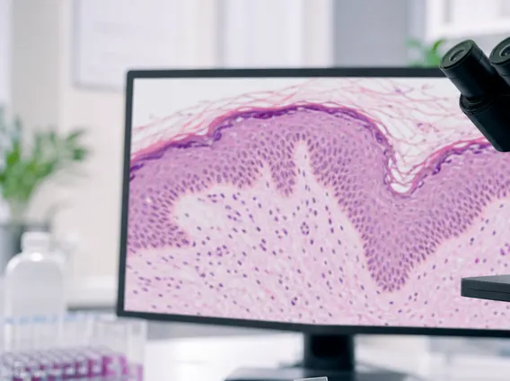
The gold standard for diagnosis is a biopsy, where tissue is obtained for microscopic examination. Pathologists identify the characteristic features of PILA, including papillary projections lined by atypical endothelial cells within dilated lymphatic channels, often with intraluminal erythrocytes and hemosiderin deposition. Immunohistochemical staining, particularly for lymphatic endothelial markers like D2-40 (podoplanin) and vascular markers like CD31 and CD34, further aids in confirming the diagnosis and differentiating it from other vascular tumors.
Treatment for Papillary Intralymphatic Angioendothelioma primarily involves complete surgical excision. Due to its locally infiltrative nature and potential for recurrence, wide local excision with clear margins is crucial. In cases where complete removal is challenging due to the tumor’s location or size, a multidisciplinary approach involving surgical oncology and possibly reconstructive surgery may be necessary. Post-operative follow-up is essential to monitor for any signs of local recurrence, which can occur even after seemingly complete removal. While recurrence is possible, metastasis is exceedingly rare, reinforcing its classification as a low-grade tumor.