Papillary Dermis
The papillary dermis is the uppermost layer of the dermis, situated directly beneath the epidermis. It plays a crucial role in skin health, providing essential nutrients to the epidermis and contributing to the skin’s overall structure and sensory functions.
Key Takeaways
- The papillary dermis is the superficial layer of the dermis, characterized by finger-like projections called dermal papillae.
- Its primary papillary dermis function includes nourishing the epidermis, housing sensory receptors, and contributing to skin elasticity.
- The papillary dermis structure is rich in thin collagen fibers, elastic fibers, capillaries, and nerve endings.
- It differs significantly from the deeper reticular dermis in terms of fiber density, organization, and primary roles.
What is the Papillary Dermis?
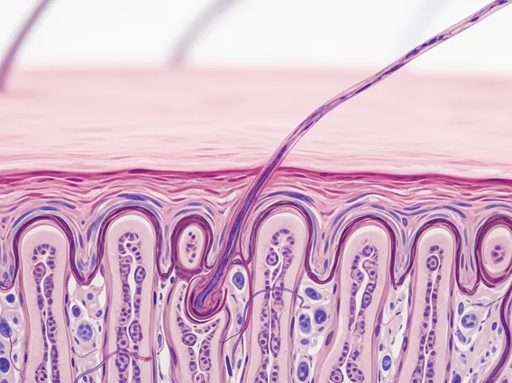
The papillary dermis is the most superficial layer of the dermis, directly underlying the basement membrane zone that separates it from the epidermis. It is named for its distinctive finger-like projections, known as dermal papillae, which interdigitate with corresponding epidermal ridges. This intricate interlocking structure significantly increases the surface area between the two skin layers, enhancing their connection and facilitating vital exchanges. This layer is critical for maintaining skin integrity and function, acting as a bridge between the avascular epidermis and the vascularized deeper dermis.
Comprising about one-fifth of the total dermal thickness, the papillary dermis is relatively thin compared to the layer beneath it. Its unique architecture is fundamental to its biological roles, ensuring the epidermis receives necessary support and nutrients. Understanding what is papillary dermis involves recognizing its anatomical position and its foundational contribution to healthy skin.
Papillary Dermis: Structure & Function
The papillary dermis structure is characterized by loose connective tissue, primarily composed of thin, loosely arranged collagen fibers (Type I and Type III) and fine elastic fibers. This arrangement allows for flexibility and resilience. Within the dermal papillae, there is an abundance of capillaries, which are tiny blood vessels responsible for delivering oxygen and nutrients to the overlying epidermis, which lacks its own blood supply. This vascular network is vital for epidermal cell metabolism and regeneration.
Beyond nutrient supply, the papillary dermis function extends to sensory perception and temperature regulation. It houses various nerve endings and sensory receptors, including Meissner’s corpuscles, which are responsible for light touch and discriminative touch sensations. The rich capillary network also plays a role in thermoregulation by allowing blood flow to increase or decrease, helping to dissipate or conserve body heat. Fibroblasts, mast cells, and macrophages are also present, contributing to tissue maintenance, immune surveillance, and wound healing processes.
Key components found within the papillary dermis include:
- Dermal Papillae: Interlocking projections that increase surface area for nutrient exchange and epidermal adhesion.
- Capillary Loops: Supply blood to the avascular epidermis.
- Nerve Endings: Detect touch, pain, and temperature.
- Collagen and Elastic Fibers: Provide strength and elasticity to the skin.
- Ground Substance: A gel-like matrix containing proteoglycans and hyaluronic acid, supporting tissue hydration and structure.
Papillary vs. Reticular Dermis
Distinguishing between the papillary and reticular layers is crucial for understanding the dermis as a whole. The papillary dermis vs reticular dermis comparison highlights their distinct structural organizations and functional specializations. While both are layers of the dermis, they differ significantly in their composition and primary roles.
The reticular dermis, located beneath the papillary dermis, is much thicker and denser. It is characterized by thick, irregularly arranged bundles of collagen fibers (predominantly Type I) and coarse elastic fibers, giving it immense tensile strength and elasticity. This layer provides the main structural support for the skin, housing larger blood vessels, nerves, hair follicles, sebaceous glands, and sweat glands. In contrast, the papillary dermis is thinner, with loosely arranged fibers, and focuses more on epidermal nourishment and sensory input.
| Feature | Papillary Dermis | Reticular Dermis |
|---|---|---|
| Location | Superficial layer, directly beneath epidermis | Deep layer, beneath papillary dermis |
| Thickness | Thinner (approx. 20% of dermis) | Thicker (approx. 80% of dermis) |
| Collagen Fibers | Thin, loosely arranged (Type I & III) | Thick, densely packed, irregularly arranged (Type I) |
| Elastic Fibers | Fine, less abundant | Coarse, more abundant |
| Primary Function | Nourishment of epidermis, light touch sensation, epidermal adhesion | Tensile strength, elasticity, houses glands and hair follicles |
| Key Structures | Dermal papillae, capillary loops, Meissner’s corpuscles | Larger blood vessels, nerves, hair follicles, sweat glands, sebaceous glands |
In essence, the papillary dermis is designed for intricate interaction with the epidermis, focusing on nutrient exchange and sensory reception, while the reticular dermis provides the bulk of the skin’s mechanical strength and houses most of its adnexal structures.