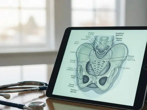
Median Umbilical Ligament
The median umbilical ligament is a fibrous cord located in the abdomen, representing a remnant of a crucial fetal structure. Understanding its anatomy and potential pathologies is important for diagnosing related abdominal conditions.
Median Umbilical Ligament
- The median umbilical ligament is a fibrous remnant of the fetal urachus, connecting the bladder to the umbilicus.
- In adults, it serves a minor role in stabilizing the bladder and is typically asymptomatic.
- Pain can arise from congenital anomalies like urachal cysts or diverticula, as well as inflammation or infection.
- Diagnosis often involves imaging techniques such as ultrasound or CT scans.
What is the Median Umbilical Ligament?
The median umbilical ligament is a fibrous cord that extends from the apex of the urinary bladder to the umbilicus. It is a vestigial structure in adults, meaning it no longer performs its original function. This ligament is a remnant of the urachus, a tube that connects the fetal bladder to the allantois, an embryonic sac involved in waste removal. After birth, the urachus normally obliterates and transforms into this non-functional fibrous band.
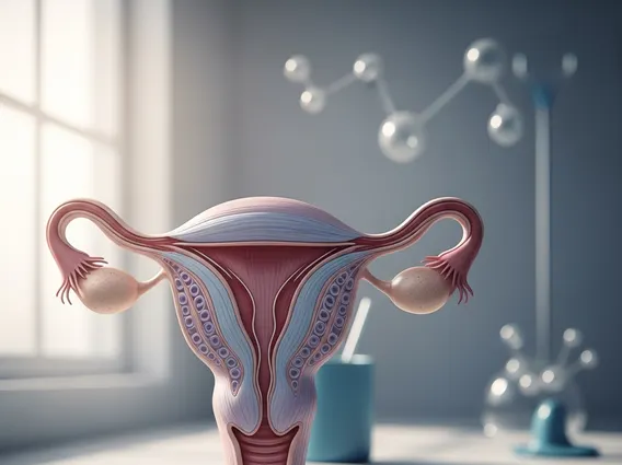
Anatomy and Function of the Ligament
The median umbilical ligament anatomy involves its course along the anterior abdominal wall, situated in the midline. It lies beneath the peritoneum, creating a raised fold on the inner surface of the abdominal wall, known as the median umbilical fold. This anatomical position places it between the two medial umbilical ligaments, which are remnants of the umbilical arteries. The primary median umbilical ligament function in adults is minimal; it contributes to the stabilization of the bladder within the pelvis, anchoring it to the abdominal wall. During fetal development, however, the urachus played a vital role in draining urine from the fetal bladder into the allantois, a function that ceases at birth with the closure of the urachus.
Causes of Median Umbilical Ligament Pain
Pain associated with the median umbilical ligament pain causes typically stem from developmental anomalies or inflammatory processes affecting the urachal remnant. While the ligament itself is usually asymptomatic, remnants of the urachus can sometimes persist, leading to various conditions. These conditions are relatively rare; for instance, symptomatic urachal anomalies are estimated to occur in less than 1 in 5,000 births.
Common causes of pain include:
- Urachal Cysts: These occur when a portion of the urachus fails to close completely, forming a fluid-filled sac. Cysts can become infected, leading to localized pain, tenderness, and fever.
- Patent Urachus: In rare cases, the urachus remains open, creating a direct connection between the bladder and the umbilicus. This can result in urine leakage from the navel and is prone to infection, causing pain and irritation.
- Urachal Diverticulum: This is an outpouching of the bladder at its apex, where the urachus attaches. It can lead to urinary stasis, infection, and subsequent pain.
- Urachitis: Inflammation of the urachal remnant, often due to infection, can cause significant pain in the lower abdomen, mimicking other acute abdominal conditions.
- Trauma or Strain: Although less common, direct trauma to the lower abdomen or severe abdominal strain can potentially irritate or inflame the ligament, leading to discomfort.
Diagnosis of these conditions often involves imaging studies such as ultrasound, CT scans, or MRI, which can visualize the urachal remnant and any associated abnormalities. Treatment typically involves antibiotics for infections and, in many cases, surgical removal of the anomalous structure to prevent recurrence and complications.