Funduscopy
Funduscopy is a vital diagnostic procedure in ophthalmology that allows healthcare professionals to examine the back of the eye. This examination provides crucial insights into ocular health and can reveal signs of various systemic conditions affecting the body.
Key Takeaways
- Funduscopy involves examining the retina, optic disc, macula, and retinal blood vessels.
- It is essential for diagnosing and monitoring eye conditions such as glaucoma, macular degeneration, and diabetic retinopathy.
- The procedure typically requires pupil dilation to achieve a clear view of the fundus.
- Beyond eye health, funduscopy can detect indicators of systemic diseases like hypertension and diabetes.
- Different techniques, including direct, indirect, and slit-lamp funduscopy, are used depending on the clinical need.
What is Funduscopy?
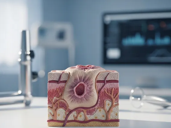
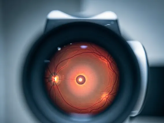
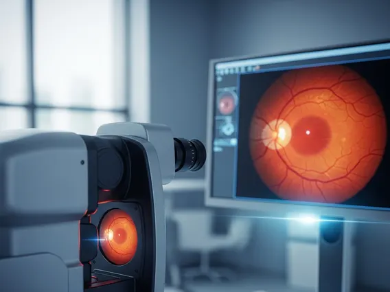
Funduscopy, also known as ophthalmoscopy, is a clinical procedure used to visualize the fundus of the eye. The fundus is the interior surface of the eye opposite the lens and includes the retina, optic disc, macula, fovea, and posterior pole, along with their associated blood vessels. This non-invasive examination allows ophthalmologists and other healthcare providers to assess the health of these critical structures, which are vital for vision. By examining the fundus, doctors can detect early signs of disease that might otherwise go unnoticed, often before symptoms become apparent to the patient.
The procedure is fundamental in routine eye care and emergency medicine. It provides a direct view of the only place in the body where blood vessels can be seen non-invasively, offering a unique window into both ocular and systemic health. The clarity and detail observed during a funduscopy can guide diagnosis, treatment planning, and monitoring of various conditions affecting the eye and beyond.
Why is a Funduscopy Exam Performed?
A purpose of funduscopy exam is primarily to diagnose and monitor a wide range of eye conditions and systemic diseases that manifest in the eye. It is a critical tool for early detection, which can significantly impact treatment outcomes and preserve vision. By carefully inspecting the retina and optic nerve, clinicians can identify subtle changes indicative of underlying health issues.
Common conditions for which a funduscopy exam is performed include:
- Diabetic Retinopathy: Damage to the blood vessels in the retina caused by diabetes.
- Hypertensive Retinopathy: Retinal damage resulting from high blood pressure.
- Glaucoma: A group of eye conditions that damage the optic nerve, often due to high pressure inside the eye.
- Macular Degeneration: Deterioration of the macula, the central part of the retina responsible for sharp, central vision.
- Retinal Detachment: A serious condition where the retina pulls away from the underlying tissue.
- Optic Nerve Disorders: Including optic neuritis or optic atrophy.
- Intracranial Pressure: Swelling of the optic disc (papilledema) can indicate increased pressure in the brain.
- Tumors: Detection of melanomas or other growths within the eye.
Regular funduscopy is particularly important for individuals with chronic conditions like diabetes and hypertension, as these diseases can cause significant damage to the retinal blood vessels before any visual symptoms appear.
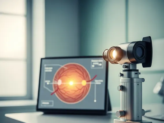
How is Funduscopy Performed?
The funduscopy procedure explained involves a systematic examination of the eye’s interior. Before the examination, the patient’s pupils are often dilated using eye drops. This step is crucial as it widens the pupil, allowing the doctor a much broader and clearer view of the fundus. The dilation process typically takes about 15-30 minutes, and patients may experience temporary light sensitivity and blurred vision afterward.
There are several methods for performing funduscopy:
| Method | Description | Key Features |
|---|---|---|
| Direct Funduscopy | Uses a handheld ophthalmoscope to shine a light into the eye and view the fundus directly. | Upright, magnified (15x) image; smaller field of view. |
| Indirect Funduscopy | Uses a bright light source (worn on the examiner’s head) and a condensing lens held in front of the patient’s eye. | Inverted, less magnified (2-5x) image; wider field of view, allowing a panoramic view of the retina. |
| Slit-Lamp Funduscopy | Performed using a slit lamp microscope in conjunction with a specialized contact or non-contact lens. | Highly magnified, stereoscopic (3D) view; excellent for detailed examination of specific areas. |
During the procedure, the patient is usually seated in a darkened room. The ophthalmologist will ask the patient to look in different directions to allow for a comprehensive view of the entire retina. The examination is generally painless, though some patients might find the bright light uncomfortable. Following the exam, patients are advised to avoid driving until the effects of the dilating drops wear off, which can take several hours.