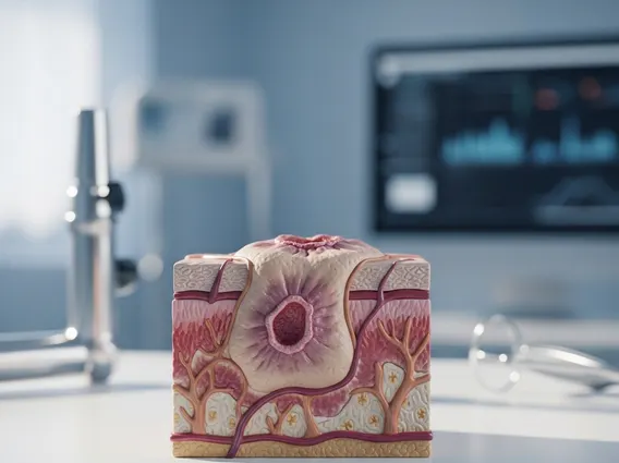
Fundoscopy
Fundoscopy is a vital diagnostic procedure used to examine the back of the eye, including the retina, optic disc, macula, and choroid. This examination provides crucial insights into ocular health and can reveal signs of systemic diseases.
Key Takeaways
- Fundoscopy is an essential eye exam for viewing the back of the eye’s internal structures.
- It helps detect various eye conditions and systemic diseases like diabetes and hypertension.
- The procedure typically involves dilating the pupils for a clearer and broader view.
- Preparation includes arranging for transportation due to temporary blurred vision and light sensitivity.
- Regular fundoscopy can aid in early diagnosis and timely intervention for maintaining eye health and overall well-being.
What is Fundoscopy: Eye Exam Explained
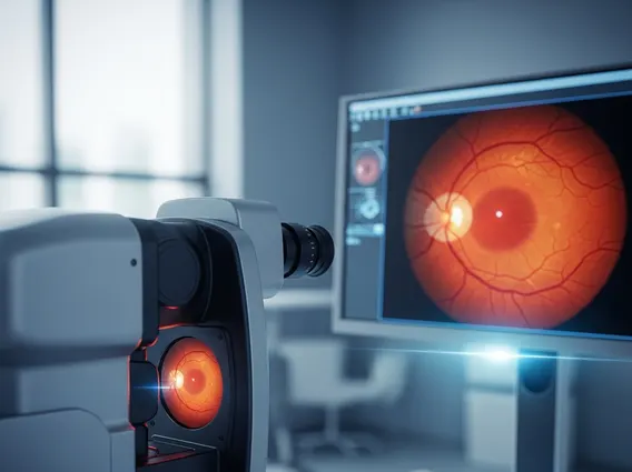
Fundoscopy refers to a diagnostic medical procedure used by ophthalmologists and other healthcare professionals to examine the posterior segment of the eye. This includes the retina, optic disc, macula, and choroid. It is also commonly known as an ophthalmoscopy. This essential eye exam allows for the direct visualization of the blood vessels, nerve tissue, and other structures at the back of the eye, which can reveal crucial information about a patient’s ocular health and overall systemic well-being.
To learn about fundoscopy examination, it’s important to understand its non-invasive nature. The procedure is typically quick and painless, providing a window into the body’s vascular and neurological health. For those seeking fundoscopy explained for beginners, think of it as a detailed internal inspection of the eye, much like looking at a photograph of the inside of your eye. It is instrumental in detecting a wide range of conditions, often before symptoms become apparent.
Conditions that can be identified through fundoscopy include:
- Diabetic retinopathy, a complication of diabetes that damages blood vessels in the retina.
- Glaucoma, characterized by damage to the optic nerve.
- Macular degeneration, a leading cause of vision loss in older adults.
- Retinal detachment, a serious condition where the retina pulls away from its supporting tissue.
- Hypertensive retinopathy, changes in the retina due to high blood pressure.
- Optic nerve damage or swelling, which can indicate neurological issues.
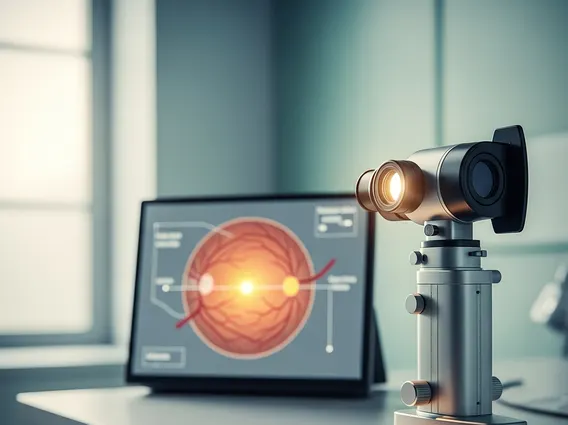
Fundoscopy Procedure and Purpose
The Fundoscopy procedure and purpose are centered around providing a clear view of the fundus, the interior surface of the eye opposite the lens. Before the examination, dilating eye drops are often administered to widen the pupils. This dilation is crucial as it allows the examiner a much broader and clearer view of the retina and optic nerve. It typically takes about 15-30 minutes for the drops to take full effect, during which time vision may become temporarily blurred and light-sensitive.
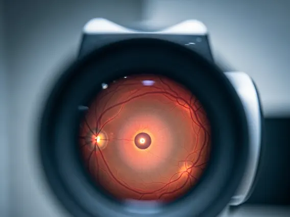
Once the pupils are dilated, the healthcare professional will use an ophthalmoscope, a specialized instrument with a light source and magnifying lenses. There are primarily two types of fundoscopy:
- Direct Ophthalmoscopy: The examiner uses a handheld ophthalmoscope close to the patient’s eye. This method provides a highly magnified, upright, but narrower view of the fundus. It is commonly used for routine eye exams.
- Indirect Ophthalmoscopy: The examiner uses a head-mounted ophthalmoscope and a separate condensing lens held in front of the patient’s eye. This method offers a wider, less magnified, inverted view of the fundus, allowing for a more comprehensive assessment of the peripheral retina.
The primary purpose of fundoscopy is early detection and monitoring of various ocular and systemic conditions. By examining the intricate network of blood vessels, the optic nerve head, and the macula, doctors can identify subtle changes that might indicate disease progression or the onset of new conditions. For instance, changes in retinal blood vessels can be an early indicator of diabetes or hypertension, while optic nerve abnormalities can signal glaucoma or neurological problems. According to the World Health Organization (WHO), regular eye examinations, including fundoscopy, are vital for preventing avoidable blindness and visual impairment, especially in populations at risk for conditions like diabetic retinopathy.
Preparing for Your Fundoscopy Exam
Preparing for your fundoscopy exam is relatively straightforward but involves a few important considerations, primarily due to the use of dilating eye drops. Patients should be aware that these drops will temporarily blur their vision and make their eyes more sensitive to light for several hours after the procedure.
Key preparation steps include:
- Arranging Transportation: It is highly recommended to have someone drive you home after your appointment, as your vision may be too impaired for safe driving.
- Bringing Sunglasses: Wearing sunglasses can significantly reduce discomfort from light sensitivity immediately after the exam.
- Informing Your Doctor: Always inform your eye care professional about any medications you are currently taking, as well as any allergies or existing medical conditions, especially glaucoma or a history of narrow-angle glaucoma, as pupil dilation may be contraindicated in some cases.
- Asking Questions: Do not hesitate to ask your doctor or the clinic staff any questions you may have about the procedure or what to expect.
Understanding these preparatory steps ensures a smoother and safer experience, allowing the eye care professional to conduct a thorough and effective fundoscopy examination.