Endometrial Biopsy
Endometrial Biopsy is a diagnostic procedure crucial for evaluating the health of the uterine lining. This article explores the procedure, its purposes, and what to expect regarding results, risks, and benefits.
Key Takeaways
- An Endometrial Biopsy involves taking a small tissue sample from the uterine lining for laboratory analysis.
- It is primarily used to investigate abnormal uterine bleeding, infertility, or to screen for uterine cancer.
- The procedure is typically quick, performed in an outpatient setting, and may cause mild discomfort.
- Results help diagnose conditions ranging from hormonal imbalances to precancerous changes or cancer.
- While generally safe, potential risks include cramping, light bleeding, infection, or, rarely, uterine perforation.
What is Endometrial Biopsy: Procedure and Purpose
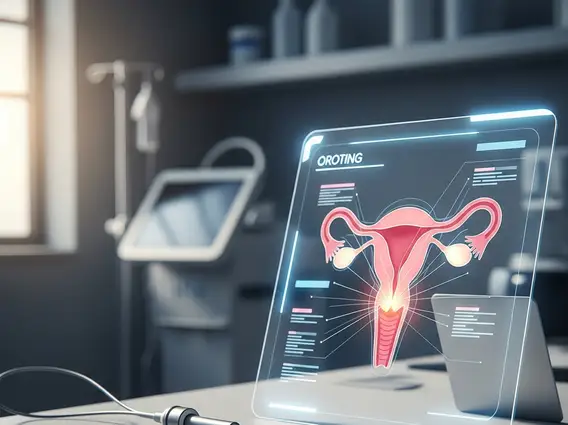
An Endometrial Biopsy refers to a medical procedure where a small sample of tissue is removed from the endometrium, the lining of the uterus. This tissue is then sent to a laboratory for microscopic examination to detect abnormalities.
The procedure, often performed in a doctor’s office, typically involves the insertion of a thin, flexible tube (pipelle) through the cervix into the uterus. The cervix may be numbed beforehand, and a speculum is used to visualize it. Suction is then applied to collect a tissue sample. Patients may experience cramping, similar to menstrual cramps, during the brief procedure, which usually lasts about 5-10 minutes.
The primary purpose of this diagnostic tool is to investigate various uterine conditions. It is frequently recommended for individuals experiencing abnormal uterine bleeding, such as heavy or prolonged periods, bleeding between periods, or postmenopausal bleeding. Other common indications include evaluating uterine health in cases of infertility, monitoring the effects of hormone therapy, or screening for endometrial hyperplasia (a precancerous condition) and uterine cancer. According to the American College of Obstetricians and Gynecologists (ACOG), it is a key tool in the workup of abnormal uterine bleeding, especially in women over 45 or those with risk factors for endometrial cancer.
Endometrial Biopsy Results: Interpretation, Risks, and Benefits
Understanding the significance of the findings from an Endometrial Biopsy is crucial for subsequent medical management. The laboratory analysis of the tissue sample can reveal several conditions. Normal results indicate healthy endometrial tissue, while abnormal findings might point to hormonal imbalances, inflammation (endometritis), endometrial polyps, hyperplasia, or cancerous cells. For instance, the presence of atypical cells often suggests a need for further investigation or treatment, as these can be precursors to malignancy. The pathologist’s report will detail the cellular characteristics, guiding the healthcare provider in formulating a diagnosis and treatment plan.
Like any medical intervention, an Endometrial Biopsy carries potential risks and benefits. Weighing these factors is important for patients considering the procedure.
Potential benefits include:
- Accurate Diagnosis: Provides definitive tissue diagnosis for various uterine conditions, including cancer, which cannot be achieved through imaging alone.
- Early Detection: Can detect precancerous changes or early-stage cancer, allowing for timely intervention and improving prognosis.
- Guidance for Treatment: Helps physicians tailor appropriate treatment plans based on specific findings, avoiding unnecessary or ineffective therapies.
- Minimally Invasive: Generally less invasive than surgical alternatives like D&C (dilation and curettage), often performed in an outpatient setting without general anesthesia.
Potential risks include:
- Pain and Discomfort: Patients commonly experience cramping, similar to menstrual cramps, during and shortly after the procedure. Over-the-counter pain relievers can help manage this.
- Bleeding and Spotting: Light bleeding or spotting is common for a few days post-procedure and usually resolves on its own.
- Infection: Though rare, there is a small risk of infection in the uterus, which may require antibiotics.
- Uterine Perforation: Extremely rare, but possible, where the instrument punctures the uterine wall. This typically requires observation or, in rare cases, surgical repair.
- Incomplete Sample: Sometimes, the tissue sample may not be sufficient for a definitive diagnosis, necessitating a repeat procedure or further diagnostic tests.
Patients should discuss any concerns about these aspects with their healthcare provider to ensure informed consent and appropriate post-procedure care, including instructions on managing discomfort and recognizing signs of complications.