Diagnosis, Screening, and Early Detection of Thymoma
Thymoma is a rare tumor that starts in the epithelial cells of the thymus gland, a small organ located behind the breastbone. Understanding the early signs, when screening and early detection make sense, and how thymoma diagnosis actually happens is central to timely, appropriate care.
Key Takeaways
- A thymoma often causes no symptoms at all, especially early on; when signs appear, they typically reflect either the tumor’s physical growth in the chest or a separate autoimmune reaction, most often myasthenia gravis.
- Thymoma is rare — per National Cancer Institute SEER registry data, roughly 0.13 diagnoses for every 100,000 people each year — so there is no population-wide screening test; most tumors are found incidentally on a chest scan done for another reason, or while investigating myasthenia gravis.
- Myasthenia gravis is the autoimmune condition most closely tied to thymoma, with the same National Cancer Institute data showing it affects roughly 30% to 65% of thymoma patients in reported case series.
- Diagnosis begins with a clinical evaluation and chest imaging — chest X-ray, CT, and sometimes MRI or PET — but only tissue examination can confirm the diagnosis and classify the tumor’s subtype.
- No blood test diagnoses thymoma directly; blood work is mainly used to confirm a suspected associated condition, such as myasthenia gravis.
Recognizing Early Thymoma Symptoms
A thymoma frequently produces no symptoms whatsoever, particularly while it is small. Two broad patterns emerge when signs are present: physical effects from the tumor’s growth inside the chest cavity, and separate signals from an autoimmune reaction the body can mount independently of how large the tumor is.
Respiratory Symptoms
As a thymoma grows, it can press on the trachea, bronchi, or lungs, causing a cough that doesn’t go away, shortness of breath, or chest pain. A hoarse voice or phrenic nerve involvement can also occur. These symptoms overlap with many common respiratory conditions, which can delay recognition. A large tumor can occasionally compress the major vein that returns blood from the upper body to the heart, producing superior vena cava syndrome — swelling of the face and neck, visibly distended neck veins, and difficulty breathing.
Neuromuscular Symptoms
The autoimmune condition most closely linked to thymoma is myasthenia gravis, a disorder in which the immune system produces antibodies against the acetylcholine receptor (and sometimes other proteins, such as titin) at the junction between nerve and muscle. According to the National Cancer Institute, in reported case series, roughly 30% to 65% of thymoma patients are also diagnosed with myasthenia gravis. Its symptoms can include:
- Drooping eyelids (ptosis)
- Double vision (diplopia)
- Difficulty swallowing or speaking
- Weakness in the arms or legs
These neuromuscular symptoms can be among the first clues that lead a doctor to look for an underlying thymoma.
Screening and Early Detection Strategies
Thymoma is uncommon: SEER registry figures compiled by the National Cancer Institute put the annual rate at roughly 0.13 diagnoses for every 100,000 people. Because the disease is so uncommon, there is no recommended population-wide screening test; testing everyone without symptoms or risk factors would mostly generate false alarms rather than catch real cases earlier.
Who Needs Screening?
There is no formal screening guideline that applies to the general population. In practice, two situations prompt a closer look: a patient already diagnosed with myasthenia gravis or another autoimmune paraneoplastic condition — since thymoma is the tumor most strongly associated with it — and a mediastinal mass discovered unexpectedly on imaging obtained for an unrelated reason. In both cases, chest imaging is used to look for a thymoma even though no dedicated screening program exists.
Current Screening Approaches
Most thymomas are picked up incidentally: a chest X-ray or CT scan ordered for a cough, chest pain, or an unrelated injury happens to reveal a mass in the mediastinum. About half of thymomas are diagnosed while the tumor is still confined within the thymus’s own outer capsule, before it has grown into surrounding tissue. Because myasthenia gravis and thymoma occur together so often, a new myasthenia gravis diagnosis is one of the more common reasons a patient ends up having chest imaging that finds a thymoma.
Thymoma Diagnosis Methods
When a thymoma is suspected, doctors combine a clinical evaluation with imaging to confirm a mass is present and understand its size, location, and relationship to nearby structures.
Initial Clinical Evaluation
The process starts with a physical exam and a review of medical history, with particular attention to respiratory symptoms, chest discomfort, and any signs of neuromuscular weakness. A thorough exam may reveal findings such as superior vena cava syndrome or neurological signs suggestive of myasthenia gravis. This initial evaluation helps guide which imaging studies come next.
Imaging Techniques
Several imaging tools are used together to locate and characterize a suspected thymoma:
- Chest X-ray: Often the first study obtained, and the way many thymomas are found in the first place, since it may be ordered for an unrelated complaint. It can suggest a mass but cannot confirm the diagnosis on its own.
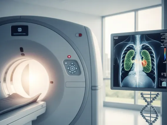
- Computed Tomography (CT) scan: The most useful imaging test for thymoma. CT with intravenous contrast helps define the tumor’s size and location and assess whether it has grown into nearby blood vessels, the sac around the heart, or the lungs.
- Magnetic Resonance Imaging (MRI): Used selectively rather than routinely — for example, to help distinguish a thymoma from normal thymus tissue or benign thymic enlargement, to evaluate whether the tumor has grown into the heart muscle, or to assess invasion of the chest wall, where it can be more informative than CT.
- Positron Emission Tomography (PET) scan: Not a routine part of initial thymoma diagnosis. It has mainly been studied in small series of thymic carcinoma, a more aggressive, related tumor type, where higher tracer uptake has been linked to more invasive disease; its overall accuracy for this purpose is still being defined.
These studies guide further diagnostic steps, including the decision about how tissue will be obtained for a definitive diagnosis.
Advanced Tests for Thymoma Confirmation
Imaging can strongly suggest a thymoma, but only examination of tumor tissue under a microscope can confirm the diagnosis and classify the tumor.
Biopsy and Histopathology
A needle biopsy, obtained under imaging guidance, can supply tissue for diagnosis before treatment is planned. When imaging strongly suggests an early-stage, confined tumor that appears fully removable, surgeons will sometimes proceed directly to surgical removal rather than a separate biopsy first — in that situation, the operation itself serves as both the diagnostic and the definitive step, since the entire tumor is available for examination.
Pathologists classify thymoma tissue using a World Health Organization system that defines several histological subtypes — A, AB, B1, B2, and B3 — based on the appearance of the tumor’s epithelial cells and how many immature lymphocytes are mixed in. A related but distinct diagnosis, thymic carcinoma, shows more clear-cut abnormal cells and lacks those immature lymphocytes. Histological subtype alone does not reliably predict how a tumor will behave; the degree to which it has invaded surrounding tissue is generally a more important factor, so pathology and imaging findings are interpreted together rather than in isolation.
Blood Tests and Biomarkers
No blood test can diagnose thymoma on its own. Blood work is more often used to support the diagnosis of an associated condition: when myasthenia gravis is suspected, a blood test for acetylcholine receptor antibodies can help confirm it, which in turn raises suspicion for an underlying thymoma. Some patients with myasthenia gravis test negative for these antibodies, so a negative result does not rule it out. Researchers continue to study other potential biomarkers for thymoma, but none is currently established for routine diagnostic use.
Frequently Asked Questions
What are the most common initial signs of thymoma?
Most people have no symptoms at all when a thymoma is first found. When symptoms occur, they are often nonspecific respiratory complaints — a persistent cough, shortness of breath, or chest pain — or neuromuscular symptoms tied to myasthenia gravis, such as drooping eyelids, double vision, or muscle weakness.
Is routine screening for thymoma recommended for everyone?
No. Thymoma is extremely rare, and there is no population-wide screening test or guideline. Most cases are found incidentally on imaging done for another reason, or through chest imaging ordered because a patient has been diagnosed with myasthenia gravis, the condition most closely associated with thymoma.
How is thymoma definitively diagnosed?
Imaging, especially a CT scan, can strongly suggest a thymoma and show its size and location, but a definitive diagnosis requires examining tumor tissue under a microscope. Depending on the situation, that tissue comes from a needle biopsy or from surgical removal of the tumor itself, which also classifies the tumor’s histological subtype.
Sources