Diagnosis, Screening, and Early Detection of Sarcoma
Sarcoma is a rare and complex group of cancers that originate in the body’s connective tissues, such as bone, muscle, fat, blood vessels, or cartilage. Understanding the process of sarcoma diagnosis, screening, and the critical role of early detection is paramount for effective treatment and improved patient outcomes.

Key Takeaways
- Sarcoma symptoms can be subtle, often presenting as a growing lump or persistent pain, so any new or changing lump deserves prompt medical evaluation.
- Accurate sarcoma diagnosis methods combine imaging, a physical exam, and a definitive biopsy with pathology and molecular testing.
- There is no standard population-wide sarcoma screening program because the disease is rare, but people with certain inherited genetic syndromes or a history of radiation exposure are followed more closely.
- Detecting sarcoma while it is still localized generally allows for less extensive treatment and a better long-term outlook than a later-stage diagnosis.
Recognizing Sarcoma Symptoms
Identifying the signs and symptoms of sarcoma can be challenging because presentation varies with the tumor’s location and type. Sarcomas can develop almost anywhere in the body, so initial signs are often non-specific and easy to mistake for more common, benign conditions. Persistent or worsening symptoms still deserve attention.
Common Warning Signs
The most common initial symptom of a soft tissue sarcoma is a painless lump or swelling that can grow over time and, if it presses on nerves or muscles, eventually cause pain or discomfort. For bone sarcomas, persistent or worsening bone pain, especially at night or with activity, is a key indicator. Other potential signs include:
- A new lump, or a lump that keeps growing, particularly if it is deep-seated or larger than about 5 centimeters (roughly the size of a lime) — a size that staging systems for soft tissue tumors treat as clinically significant.
- Pain or tenderness in the affected area that may become more noticeable as the tumor grows.
- Swelling or limited movement in a limb or joint.
- An unexplained bone fracture, especially if it is accompanied by pain.
- Abdominal pain, nausea, or a change in bowel habits if the sarcoma is in the abdomen.
Sarcomas overall are rare: soft tissue sarcomas alone make up well under 1% of new cancer diagnoses in the United States each year, according to National Cancer Institute (NCI) data, which is part of why these subtle signs are easy to overlook.
When to Consult a Doctor
It is important to see a doctor for any persistent or concerning symptom, especially a new or growing lump, unexplained pain, or swelling that does not resolve. Most lumps and aches are not sarcoma, but a medical evaluation is the only way to rule it out. Earlier consultation allows for prompt investigation, which can shorten the diagnostic timeline and widen the treatment options available. Do not delay seeking medical advice if warning signs are worsening or occur alongside unexplained weight loss or fatigue.
How Sarcoma is Diagnosed
Diagnosing sarcoma is a multi-step process that combines a clinical assessment, imaging, and definitive tissue analysis to confirm the diagnosis and identify the specific sarcoma subtype. These sarcoma diagnosis methods guide every treatment decision that follows.
Imaging and Physical Exams
Evaluation typically starts with a physical exam, where the doctor checks the size, tenderness, and mobility of the lump or painful area. Imaging then helps visualize the suspected tumor:
- X-rays: Often the first step for bone pain or a suspected bone tumor.
- Ultrasound: Can help distinguish a solid mass from a fluid-filled cyst in soft tissue.
- Magnetic Resonance Imaging (MRI): Provides detailed soft-tissue images, useful for defining tumor size, depth, and closeness to nearby structures.
- Computed Tomography (CT) scans: Used to assess the tumor’s extent, especially in the chest or abdomen, and to check for spread.
- Positron Emission Tomography (PET) scans: Can help identify metabolically active tissue, useful for staging and finding distant spread.
These studies help the care team plan the safest and most informative way to biopsy the tumor.
Biopsy and Pathology
A biopsy — removing a small sample of the suspicious tissue for a pathologist to examine — is the only way to confirm sarcoma and identify its specific type. The type of biopsy used generally depends on the tumor’s size and location:
- Core needle biopsy: A wide needle removes several tissue samples, often guided by ultrasound, CT, or MRI.
- Incisional biopsy: A surgeon cuts out a portion of the tumor for testing, an option chosen if a needle sample would be unsafe to obtain or came back inconclusive.
- Excisional biopsy: The entire lump is removed, an approach usually reserved for smaller, superficial masses.
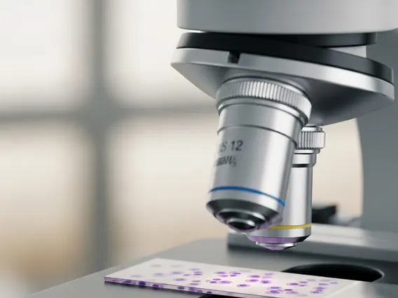
A pathologist experienced with sarcoma then examines the tissue under a microscope and may run additional tests — such as immunohistochemistry, cytogenetic analysis, or molecular profiling — to classify the exact subtype, since different sarcoma subtypes respond differently to treatment. Careful planning of the biopsy matters, since a poorly placed one can complicate the surgery that follows.
Sarcoma Screening and Risk Factors
Because sarcoma is rare, there is no evidence-based screening test recommended for the general population. Instead, closer monitoring is generally reserved for people with specific risk factors that raise their likelihood of developing the disease.
Who Should Be Screened?
Most sarcomas occur without any known cause, but certain inherited conditions and past exposures raise the risk, including:
- Inherited genetic syndromes: Li-Fraumeni syndrome, neurofibromatosis type 1 (NF1), Gardner syndrome, and Werner syndrome are each linked to a higher risk of soft tissue sarcoma; hereditary retinoblastoma is separately linked to a higher risk of osteosarcoma, a bone sarcoma.
- Previous radiation therapy: Radiation given for an earlier cancer can raise the risk of a sarcoma developing years later in the treated area.
- Long-standing lymphedema: Chronic swelling in an arm or leg — including lymphedema that can follow lymph node removal or radiation for breast cancer — is a recognized risk factor for a rare subtype called angiosarcoma.
People with one of these risk factors may be offered more frequent clinical exams and, in some cases, periodic imaging to help catch a new tumor early.
Current Screening Approaches
For the general population, there is no routine screening test for sarcoma the way there is for breast or colorectal cancer; awareness of symptoms and prompt evaluation remain the main path to an early diagnosis. NCI notes that a formal screening test for the general population does not currently exist. For people identified as high-risk, follow-up may still include regular clinical exams and, depending on the specific risk factor, periodic imaging of the affected area. For instance, someone with NF1 might have closer surveillance of existing neurofibromas, since a small number can transform into a malignant peripheral nerve sheath tumor.
The overall approach favors individualized risk assessment and tailored follow-up rather than broad population-based screening.
The Importance of Early Detection
Because sarcomas can grow and spread if left untreated, catching one at an early stage can change the entire course of treatment, offering more options and generally a better prognosis.
Impact on Treatment Success
When sarcoma is found before it has grown large or spread, surgery to remove it — the main treatment for most sarcomas — is often more straightforward. Diagnosing the tumor early can also allow for a less extensive operation, which may help preserve a limb or organ that a larger tumor would put at risk. When the cancer is caught early, the need for additional therapies such as radiation or chemotherapy may also be lower, though this depends on the specific tumor type and grade. As with most cancers, a sarcoma diagnosed while still localized tends to carry a better outlook than one found after it has spread; the specific survival statistics by stage are covered in a dedicated overview rather than repeated here.
Improving Patient Outcomes
Beyond the immediate treatment, detecting sarcoma early can mean a lower risk of recurrence, less time spent on aggressive treatment, and a faster return to daily activities. For many patients, that translates into better physical function and a reduced psychological burden during recovery. Because outcomes vary widely by sarcoma subtype, tumor grade, and how far the disease has spread, anyone wanting the specific survival figures for their own situation should discuss them directly with their care team.
Frequently Asked Questions
What are the first signs of sarcoma?
The earliest sign is often a new, painless lump or swelling that gradually grows. For bone sarcomas, persistent or worsening bone pain, especially at night, is a common early symptom. Swelling, tenderness, or limited movement in the affected area can also occur. Because these symptoms are easy to mistake for benign conditions, any persistent or worsening change is worth a prompt medical evaluation.
Can sarcoma be cured if caught early?
Sarcoma found while still localized, meaning it has not spread beyond where it started, can often be treated successfully, and complete surgical removal is the foundation of treatment for many sarcomas. Radiation or chemotherapy may also be used to lower the chance of recurrence. There is no guarantee of a cure for any individual case, but confinement to the original site generally gives the best chance of a good outcome.
Is there a blood test for sarcoma?
No single blood test can reliably diagnose sarcoma. Blood tests may be used to check general health or organ function, but they are not specific enough to identify sarcoma on their own. Diagnosis relies on a physical exam, imaging such as MRI or CT, and a biopsy with pathology review of the tissue. Research into potential biomarkers is ongoing, but none are yet part of routine diagnostic protocols.
Sources
- National Cancer Institute – Soft Tissue Sarcoma Treatment (PDQ®)–Patient Version, 2025
- National Cancer Institute – Soft Tissue Sarcoma Treatment (PDQ®)–Health Professional Version, 2025
- National Cancer Institute – Osteosarcoma and Undifferentiated Pleomorphic Sarcoma of Bone Treatment (PDQ®)–Patient Version, 2025
- National Cancer Institute, SEER Program – Cancer Stat Facts: Soft Tissue Including Heart Cancer