Ct Angiography
Computed Tomography (CT) Angiography is a sophisticated medical imaging technique that provides detailed views of blood vessels throughout the body. It plays a crucial role in diagnosing various vascular conditions by highlighting the structure and flow within arteries and veins.
Key Takeaways
- Computed Tomography (CT) Angiography uses X-rays and a contrast dye to create detailed images of blood vessels.
- The procedure involves injecting a contrast agent, followed by a rapid CT scan to capture images as the dye flows through the vessels.
- It is a non-invasive method for detecting conditions like aneurysms, blockages, and narrowing in arteries and veins.
- Benefits include high diagnostic accuracy, speed, and the ability to visualize complex vascular structures.
- CT angiography is vital for planning treatments for cardiovascular diseases and other vascular disorders.
What is Computed Tomography (CT) Angiography?
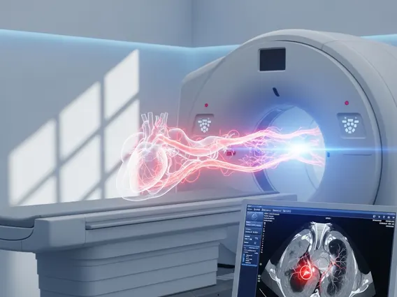
Computed Tomography (CT) Angiography is a diagnostic imaging test that combines a CT scan with an injection of a special dye, known as a contrast agent, to produce detailed images of blood vessels and blood flow. This advanced technique allows medical professionals to visualize arteries and veins in various parts of the body, including the brain, heart, lungs, kidneys, and limbs. By highlighting the blood vessels, CT angiography helps identify abnormalities that might not be visible on a standard CT scan or X-ray.
The primary purpose of CT angiography is to detect and assess conditions affecting the vascular system. It is particularly effective in identifying blockages, narrowings (stenosis), aneurysms (bulges in vessel walls), and other structural issues that can impede blood flow or pose a risk of rupture. This non-invasive method provides cross-sectional images that can be reconstructed into 3D views, offering a comprehensive understanding of the vascular anatomy and pathology.
The CT Angiography Procedure Explained
The process of a CT angiogram typically begins with preparation, which may include fasting for a few hours before the scan and discussing any allergies, especially to iodine-based contrast agents. During the procedure, the patient lies on a motorized table that slides into a large, donut-shaped CT scanner. An intravenous (IV) line is inserted, usually into a vein in the arm, through which the contrast dye is injected.
As the contrast agent travels through the bloodstream, it makes the blood vessels appear bright on the CT images. The CT scanner then rapidly takes multiple X-ray images from different angles. This rapid acquisition of images is crucial to capture the dye as it flows through the target vessels, providing a dynamic view of the vascular system. Patients are often asked to hold their breath briefly during the scan to minimize motion artifacts and ensure clear images. The entire scanning process is typically quick, often lasting only a few minutes, though the total appointment time may be longer due to preparation and post-scan observation.
Uses and Benefits of CT Angiography
CT angiography is a versatile diagnostic tool with numerous applications across various medical specialties. Its ability to provide high-resolution images of blood vessels makes it invaluable for diagnosing and managing a wide range of vascular conditions. For instance, it is frequently used to detect coronary artery disease by identifying plaque buildup in the arteries supplying the heart, which is a leading cause of heart attacks. According to the World Health Organization (WHO), cardiovascular diseases are the leading cause of death globally, underscoring the critical need for accurate diagnostic tools like CT angiography.
The benefits of CT angiography extend beyond cardiac imaging. It is also instrumental in diagnosing conditions such as:
- Aortic aneurysms: Detecting and monitoring bulges in the body’s largest artery.
- Pulmonary embolism: Identifying blood clots in the lung arteries.
- Peripheral artery disease: Locating blockages in the arteries of the legs and arms.
- Renal artery stenosis: Diagnosing narrowing of the arteries supplying the kidneys, which can cause high blood pressure.
- Cerebral aneurysms and stroke: Assessing blood vessels in the brain to identify risks or causes of stroke.
Compared to traditional angiography, which is more invasive, CT angiography offers a less invasive alternative with a lower risk of complications. It also provides detailed 3D reconstructions, aiding surgeons and interventional radiologists in planning procedures such as stent placement or surgical repair. The speed of the scan is another significant advantage, especially in emergency situations where rapid diagnosis is critical.